| Journal of Current Pharmacology, ISSN 0000-0000 online, Open Access |
| Article copyright, the authors; Journal compilation copyright, J Curr Pharmacol and Elmer Press Inc |
| Journal website https://jcp.elmerpub.com |
Original Article
Volume 1, July 2026, e1

Apigenin Mitigates Neurochemical Alterations, Oxidative and Nitrergic Stress Induced in Two-Hit Neurodevelopmental Model of Schizophrenia in Rats
Adeniyi Nicholas Adesoyea, Yusuf Usmanb, c  , Adewale Ganiyu Bakrea, c , Wakeel Adegokea, Rotimi Olatundea, Adegbuyi Oladele Aderibigbea
aDepartment of Pharmacology and Therapeutics, Faculty of Basic Medical Sciences, College of Medicine, University of Ibadan, Ibadan, Oyo State, Nigeria
bDepartment of Pharmacology, Faculty of Basic Medical Sciences, College of Medicine, Federal University of Health Sciences Ila-Orangun, Ila-Orangun, Osun State, Nigeria
cCorresponding Authors: Adewale Ganiyu Bakre, Department of Pharmacology and Therapeutics, Faculty of Basic Medical Sciences, College of Medicine, University of Ibadan, Ibadan, Oyo State, Nigeria; Yusuf Usman, Department of Pharmacology, Faculty of Basic Medical Sciences, College of Medicine, Federal University of Health Sciences Ila-Orangun, Ila-Orangun, Osun State, Nigeria
Manuscript submitted January 10, 2026, accepted March 28, 2026, published online July 13, 2026
Short title: Apigenin Mitigates Schizophrenia Phenotypes
doi: https://doi.org/10.14740/jcp1
| Abstract | ▴Top |
Background: Schizophrenia is a complex neurodevelopmental disorder arising from the interaction of genetic and environmental factors across critical developmental periods. Oxidative stress and dysregulated neurotransmission are central mechanisms implicated in its pathophysiology. Apigenin, a naturally occurring flavonoid, possesses notable antioxidant and neuromodulatory properties, suggesting potential therapeutic value as a broader-spectrum alternative to conventional antipsychotic agents. This study evaluated the efficacy of apigenin in attenuating behavioral and neurobiochemical alterations induced by isolation rearing combined with chronic ketamine exposure in two-hit neurodevelopmental model of schizophrenia.
Methods: Sixty-four (N = 64) male Sprague–Dawley rats weaned on postnatal day (PND) 21 were assigned to either group housing or isolation rearing from PND 23. Animals were further subdivided into treatment groups receiving saline (0.9% NaCl) or ketamine (16 mg/kg, i.p.) from PND 28 to 85. From PND 60 to 85, selected groups received apigenin (8 mg/kg, i.p.) or risperidone (0.5 mg/kg, i.p.) as interventions. Behavioral assessments using the open field test (OFT) and forced swim test (FST) were conducted, followed by biochemical and histopathological analyses of the prefrontal cortex, hippocampus, and amygdala.
Results: Apigenin significantly reduced hyperlocomotion and immobility in behavioral tests, biochemically, reduced oxidative stress by lowering malondialdehyde and nitrite levels, enhanced antioxidant enzyme activities, normalized dopaminergic transmission, and reduced acetylcholinesterase activity. Histological findings revealed restoration of neuronal density and structural integrity across examined brain regions.
Conclusion: Apigenin demonstrated broad therapeutic potential by mitigating behavioral abnormalities, oxidative stress, and neurochemical disturbances relevant to the pathogenesis of schizophrenia.
Keywords: Apigenin; Schizophrenia; Two-hit; Neurodevelopmental; Neurochemical; Oxidative stress; Nitrergic; Rats
| Introduction | ▴Top |
Schizophrenia is a severe neuropsychiatric disorder that affects approximately 1% of the global population and constitutes a major contributor to the global burden of disease despite its relatively low prevalence [1]. The disorder is characterized by a heterogeneous symptoms pattern, broadly classified into positive symptoms such as hallucinations, delusions, and disorganized speech or behavior, negative symptoms including social withdrawal, reduced motivation, and diminished emotional expression, as well as cognitive impairments involving deficits in working memory and information processing [2]. Schizophrenia is predominantly managed through pharmacological interventions [3, 4], specifically the administration of antipsychotic medications that modulate neurotransmitter systems, most notably dopaminergic pathways [5]. While these agents are highly effective at attenuating positive symptoms, they demonstrate limited efficacy in addressing negative and cognitive symptoms [6].
The onset of schizophrenia typically occurs during late adolescence or early adulthood [7] and its etiology reflects a complex interplay among genetic vulnerability, neurotransmitter dysregulation, and environmental influences [8]. Importantly, interactions between genetic and environmental risk factors during critical periods of brain development, particularly the prenatal and early postnatal stages, play a significant role in shaping disease onset and progression [9]. Schizophrenia manifests differently in men and women [5]. Men are more likely to be diagnosed earlier than women, with men typically experiencing an earlier onset of the disorder, more pronounced negative symptoms, and poorer functioning in the early stages of the illness [3, 5].
Environmental stressors have been well established as major triggers in the development of schizophrenia [10]. Early-life stressors, such as maternal separation, as well as stress experienced during adolescence or adulthood, have been shown to induce oxidative stress [5] and contribute to circuit-level dysregulation that disrupts brain maturation and connectivity during sensitive neurodevelopmental windows [11]. Oxidative stress arising from excessive production of reactive oxygen species (ROS) and reactive nitrogen species (RNS) or from impaired endogenous antioxidant defenses has emerged as a central mechanism in the pathogenesis of schizophrenia [12–14].
Alterations in neuronal transmission are fundamental to the pathophysiology of schizophrenia, with dopamine (DA), serotonin, glutamate, and gamma-aminobutyric acid (GABA) identified as key neurotransmitter systems involved in the disorder [1]. Notably, glutamate receptor antagonists induce behavioral and molecular changes that closely resemble schizophrenia-like phenotypes [5]. Substantial evidence indicates that inhibition of N-methyl-D-aspartate (NMDA) receptors located on inhibitory interneurons results in an excitatory–inhibitory imbalance within cortical circuits [15]. Postmortem studies further demonstrate microstructural and functional abnormalities in these circuits in individuals with schizophrenia [16]. Dysfunction of the cholinergic system has also been implicated in the clinical manifestations of schizophrenia [17]. The cholinergic system plays a critical role in modulating cognition and emotional regulation, and disruption of these functions contributes to schizophrenia-related symptoms [18]. Moreover, acetylcholine release modulates dopaminergic activity in the striatum, highlighting a close functional interaction between cholinergic and dopaminergic neurotransmission [18, 19]. Abnormal cholinergic signaling in schizophrenia may therefore exert its effects partly through altered DA release, potentially mediated by changes in acetylcholinesterase (AChE) activity [20–22].
Chronic administration of ketamine, a dissociative NMDA receptor antagonist, at sub-anesthetic doses induces glutamatergic hypofunction, dopaminergic dysregulation, increased AChE activity, and redox imbalance through the generation of oxidative stress in animal models, mechanisms that are strongly implicated in the pathogenesis of schizophrenia [23, 24]. Consequently, both acute and chronic sub-anesthetic ketamine exposure has been widely employed as a pharmacological tool for modeling schizophrenia-like phenotypes in rodents [25].
Neurodevelopmental animal models of schizophrenia employ a range of approaches, including genetic manipulation, prenatal insults, pharmacological interventions, and stress-based protocols during postnatal development, to recapitulate key features of the disorder [26]. Two-hit rodent models, which combine environmental risk factors at different developmental stages (prenatal, early postnatal, adolescence, or adulthood) with pharmacologically induced neurochemical disturbances, have been particularly valuable for elucidating schizophrenia etiology [27, 28]. For instance, the combination of social isolation rearing, a well-established environmental risk factor, with chronic ketamine administration has been shown to more accurately capture the complexity of schizophrenia pathology within a two-hit framework [6]. In such neurodevelopmental models, redox imbalance is a prominent feature, particularly in social isolation paradigms that examine the impact of postnatal social deprivation on brain development, leading to schizophrenia-like behavioral and neurobiological traits [12].
The combined exposure to early-life environmental stress and glutamate receptor antagonism exerts synergistic detrimental effects on behavior, as well as on molecular and cellular neurodevelopmental processes [5]. Accordingly, two-hit animal models have been instrumental in advancing understanding of how early-life stress increases vulnerability to psychiatric disorders later in life [29].
Apigenin is a flavone belonging to the broader class of flavonoids, a diverse group of naturally occurring polyphenolic compounds synthesized by plants [30]. It is abundantly present in vegetables such as spinach, tomatoes, onions, and celery; fruits including cherries, apples, grapes, and oranges; herbs such as chamomile, thyme, and peppermint; and plant-based beverages including tea, beer, and wine [31]. Apigenin has been widely reported to exhibit potent antioxidant, anti-inflammatory, and anticancer properties [32]. Numerous studies demonstrate its ability to scavenge free radicals, reduce oxidative stress, and inhibit pathological processes associated with various diseases [31]. Also, apigenin inhibition of AChE in amnesic mice to demonstrate the protective effect of apigenin in Alzheimer’s disease has been reported [33–35]. Furthermore, apigenin supplementation in rodents has also shown increased sedation or reduced locomotor activity in sleep improvement study [30, 33].
This study aimed to evaluate the efficacy of apigenin as a natural agent in mitigating the underlying neuropathological conditions: neurochemical alterations, as well as oxidative and nitrergic stress that trigger the onset and progression of schizophrenia symptoms modeled by the two-hit neurodevelopment hypothesis.
| Materials and Methods | ▴Top |
Drugs and chemicals
Ketamine hydrochloride (Sigma-Aldrich, St. Louis, MO, USA), apigenin (Sigma-Aldrich, St. Louis, MO, USA), risperidone (Sigma-Aldrich, St. Louis, MO, USA), dimethyl sulfoxide (DMSO) (Sigma-Aldrich, St. Louis, MO, USA), 5,5-dithiobis-(2-nitrobenzoic acid) (DTNB), thiobarbituric acid (TBA) (0.375%), Griess reagents; sulfanilamide, N-(1-naphthyl) ethylenediamine dihydrochloride (NEDD), sodium nitrite (NaNO2), hydrochloric acid (HCl), n-butanol, phosphate-buffered saline (PBS), ethanol, phosphate buffer (pH 7.4), hydrogen peroxide (H2O2), specific enzyme-linked immunosorbent assay (ELISA) kits (BioLegend, San Diego, USA), Biuret’s reagents, sulfuric acid, acetone, and distilled/deionized water were used. All chemicals/reagents used were of analytical grades (Sigma-Aldrich, St. Louis, MO, USA).
Experimental animal
Male Sprague–Dawley rats (21 days old) were obtained from central animal house, College of Medicine, University of Ibadan and kept in plastic cages in a controlled environment (25 ± 1 °C, 12:12 light/dark cycle, 40–70% relative humidity) with unlimited access to standard rodent pellet feed and water ad libitum throughout the experimental period. The animals were acclimatized for 1 week and randomly assigned into groups according to the experimental design. All treatments and experimental procedure were in strict compliance with the National Institute of Health Guide for Care and Use of Laboratory Animals (Publication No. 85-23 revised 1985).
Dosage and drug preparation
Apigenin (8 mg/kg) dosage was based on preliminary results obtained from ketamine-induced stereotypy behavior. Doses of ketamine (16 mg/kg), saline (0.9% NaCl), and risperidone (0.5 mg/kg) administered were selected as previously established [6, 24]. Apigenin solution was constituted in 5% DMSO (Vehicle). Risperidone was dissolved and diluted with distilled water, whereas, ketamine was diluted in 0.9% saline [23].
Experimental protocol
Effect of apigenin on schizophrenia-like phenotypes induced by double-hit neurodevelopmental model in rats was evaluated as previously described with little modifications [6]. At post-weanling period (postnatal day (PND) 23), the animals were assigned to either group housing rearing (eight animals per cage) cohort or social isolation housing rearing (each of the eight animals housed individually). From PND 28, a repeated single dose of ketamine (16 mg/kg, i.p) or equivalent volume of saline (0.9 % NaCl) was administered intraperitoneally three times a week (Monday, Wednesday, and Friday) to either of the group till PND 56 [36]. Thereafter on PND 57, the routine administration of ketamine or saline was reduced to two times a week (Monday and Friday) until PND 85. From PND 60 to 85, apigenin (8 mg/kg) or risperidone (0.5 mg/kg) was introduced as intervention to animals post ketamine administration. Thirty (30) minutes after apigenin or risperidone administration on PND 81 and 84, behavioral tests, open field test (OFT) (PND 81), and forced swim test (FST) (PND 84) were conducted to assess the effect of apigenin/risperidone on induced hyperlocomotion and behavioral despair, respectively. Twenty-four (24) hours after the last treatment (PND 86), the animals were sedated using ethyl ether and sacrificed by cervical dislocation. Brain samples were immediately collected for biochemical and histological analyses.
Animal grouping and treatments
Sixty-four (N = 64) male Sprague–Dawley rats (21 days old) were randomly divided into eight groups (n = 8) as follows: group 1: group housed and received saline (10 mL/kg, i.p. GHV); group 2: isolation housed and received saline (10 mL/kg, i.p. ISV); group 3: group housed and received ketamine (16 mg/kg, i.p.) (GHK); group 4: isolation housed and received ketamine (16 mg/kg, i.p.) (ISK); group 5: group housed and received ketamine (16 mg/kg, i.p.) injected + apigenin (8 mg/kg, i.p.) (GHKAPG); group 6: isolation housed and received ketamine (16 mg/kg, i.p.) + apigenin (8 mg/kg, i.p.) (SIKAPG); group 7: group housed and received ketamine (16 mg/kg, i.p.) + risperidone (0.5 mg/kg, i.p.) (GHKRISP); and group 8: isolation housed and received ketamine (16 mg/kg, i.p.) + risperidone (0.5 mg/kg, i.p.) (SIKRISP).
Behavioral assessment
1) Open field test
Locomotor activity was assessed on PND 81 using the OFT to evaluate the antipsychotic-like effects of apigenin [37]. The apparatus consisted of a square activity cage (28 × 28 × 28 cm) with three opaque sides and one transparent Plexiglas side. The floor was marked with 16 equal squares (7 × 7 cm each) using black paint. Thirty minutes after treatment, rats were individually placed in the center of the box and allowed to explore for 5 min. Locomotor activity was quantified as the number of squares crossed with all four paws, recorded by an independent observer. After every trial, the apparatus was cleaned up with 70% ethanol and dried.
2) Forced swim test
The FST was conducted to assess despair-like behavior, predictive of negative symptoms in psychosis [38]. On PND 84, each rat was placed individually in a transparent glass cylinder (46 cm height × 20 cm diameter) filled with water to a depth of 30 cm at 25 °C. Rats were forced to swim for 6 min, and immobility time was recorded during the last 5 min using a stopwatch, discarding the first minute of escape-directed activity [39]. After testing, animals were gently dried and returned to a warmed, open space until fully dry [38]. Reduction in immobility was interpreted as an index of antidepressant-like activity [40].
3) Tissue preparation
Following behavioral assessments, harvested brain samples of three (3) animals per group were dissected into amygdala, hippocampus, and prefrontal cortex (PFC), and each region was placed in ice-cold phosphate-buffered solution (pH 7.4). Tissues were minced, homogenized using a Teflon homogenizer, and centrifuged at 10,000 × g for 10 min at 4 °C. The resulting supernatants were collected as mitochondrial fractions (PMF) for biochemical assays.
Determination of each section’s brain oxidative and nitrergic stress markers
1) Glutathione assay
Determination of reduced glutathione (GSH) concentration was based on the protocol of Moron et al (1979) [41]. This involved the reaction of sulfhydryl compounds with DTNB to form a characteristic yellow product that corresponds to the concentration of GSH in the sample. The absorbance was read against blank with a spectrophotometer at a wavelength of 412 nm within 30 min of color development. The reduced GSH concentration in each brain sample was expressed in micromoles per gram tissue.
2) Lipid peroxidation assay
Determination of lipid peroxidation was done by the method of Ohkawa et al (1979) [42]. The principle is based on the reaction of malondialdehyde (MDA) with TBA to form a characteristic pink complex whose optical density corresponds to the concentration of MDA. The supernatant was collected and 0.9 mL was added to 0.1 mL TBA (0.375%). Resulting mixture was read against blank in a spectrophotometer at a wavelength of 532 nm. The extinction coefficient E532 nm used for quantifying TBARS was 1.56 × 10⁵ M−1 cm−1. The MDA content of the samples was expressed in nanomole of MDA per milligram protein (nmol/mg protein).
3) Nitrite assay
Determination of nitrite (NO) levels was carried out by method of Green et al (1982) [43]. The NO estimation in tissue sample was based on the principle that NO forms colored azo dye complexes when reacted with primary aromatic amines in acidic solution, and the colorimetric absorbance of the diazonium salt-colored complexes corresponds to the measure of NO levels. Griess reagent of 0.5 mL was added to 0.5 mL of brain supernatant aliquot. The resulting solution was incubated for 10 min at room temperature in the dark and the absorbance was measured with a spectrophotometer at a wavelength of 540 nm. The brain NO levels of supernatants were expressed as µmol/mg protein.
4) Superoxide dismutase assay
Superoxide dismutase (SOD) activity was measured by the Misra and Fridovich (1972) method [44]. Superoxide (O2•−) radicals are produced when adrenaline is oxidized, and these radicals are then converted to colored products called adrenochromes as a result of inhibition by SOD. The absorbance was measured using a spectrophotometer at a wavelength of 480 nm. The specific activity of SOD was reported as units per mg of protein.
5) Estimation of AChE activity
AChE activity was measured using protocol described by Ellman et al (1961) [45]. This method was founded on the idea that AChE hydrolyzes acetylthiocholine iodide into two main products: acetic acid and thiocholine. These products react with DTNB to form a distinctive yellow complex, the optical density of which was measured calorimetrically. A yellow color developed and at 412 nm, the absorbance was measured. AChE activity was expressed in nanomole of acetylthiocholine iodide hydrolyzed per mg of protein per minute.
6) Protein assay
Estimation of protein in test samples used the biuret method as described by Gornall et al (1949) [46]. The biuret reagent was formed by dissolving 3 g of CuSO4.5H2O in 500 mL of 0.2 M NaOH, adding 5 g of potassium iodide, and then adding 0.2 M NaOH to make up to 1 L. Following the biuret reagent preparation, the brain samples were added to biuret reagent in triplicate. The mixture was incubated for 30 min at room temperature, and the absorbance was measured at 540 nm. The protein content of the sample was extrapolated from the standard curve using bovine serum albumin as standard.
7) Determination of DA concentrations
Brain levels of DA were determined in the amygdala, PFC, and hippocampus using ELISA according to manufacturer’s instructions. The concentration of DA was expressed as ng/g tissue.
Histological assessment
1) Hematoxylin and eosin staining
The post-fixed brains (n = 3) were processed for histological findings in the amygdala, hippocampus, and the PFC. Multiple serial sections of the brain were collected by cutting coronal brain sections of the amygdala, hippocampus, and PFC at 25 mm using a microtome. Then the collected serial sections were further subjected to slide preparation and mounting process which sequentially includes dehydration, clearing, infiltration, embedding, sectioning, and lastly staining with hematoxylin and eosin (H&E) dye. All of the prepared slides were properly labeled to identify their corresponding treatment groups and were viewed with a binocular microscope at × 400 magnification for morphological architecture and structural integrity of the amygdala, hippocampal, and PFC brain regions. Photomicrographs of each section under view were taken with the aid of a digital camera and the images were carefully assessed for structural alterations. Furthermore, the neuronal densities of cells in these brain regions were assessed by counting the total viable and functional cells characterized by round shape with intact cytoplasmic membrane and no distortion of nuclear materials.
Statistical analysis
The data were expressed as mean ± standard error of mean (SEM) and analyzed using one-way analysis of variance (ANOVA) and Tukey’s post hoc test for multiple comparison where appropriate using Graphpad Prism version 4 (San Diego, CA, USA). A level of P < 0.05 was considered statistically significant for all tests.
| Results | ▴Top |
Apigenin attenuates ketamine-induced hyperlocomotion on weanling rats in two-hit model of neurodevelopmental psychosis
Ketamine significantly increased locomotor activity in group-housed rats (F(3,16) = 10.89, P < 0.0004) compared with vehicle-treated controls and reduced locomotor activity in isolated rats compared with vehicle-treated isolated controls (F(3,16) = 56.71, P < 0.0001). Apigenin and risperidone significantly reduced locomotor activity relative to the ketamine-only group (P < 0.0004) in group-housed rats. The results are depicted in Figure 1.
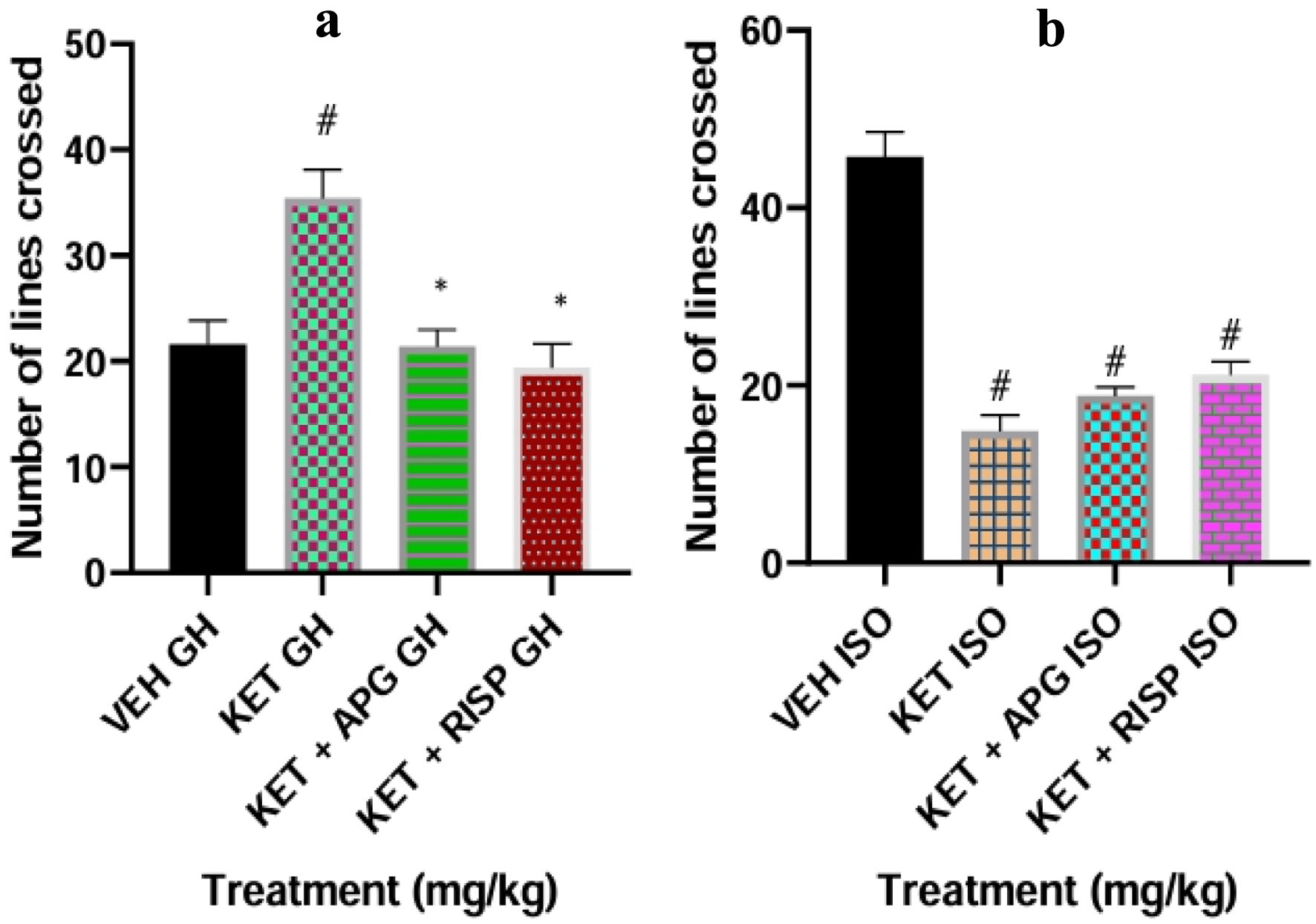 Click for large image | Figure 1. Effect of APG on locomotor activity of weanling rats subjected to two-hit model of neurodevelopmental psychosis in OFT. (a) Group-housed animals. (b) Isolated-rearing animals. Bar represents the mean ± SEM of eight animals. *P < 0.05 as compared with KET GH/KET ISO followed by ANOVA and Tukey’s post hoc test for multiple comparisons. #P < 0.05 as compared with VEH GH/VEH ISO followed by ANOVA and Tukey’s post hoc test for multiple comparisons. APG: apigenin; ANOVA: analysis of variance; GH: group-housed; ISO: isolated; KET: ketamine; OFT: open field test; RISP: risperidone; SEM: standard error of the mean; VEH: vehicle. |
Effect of Apigenin on immobility in FST on weanling rats subjected to two-hit model of neurodevelopmental psychosis
Ketamine increased immobility in group-housed rats compared with vehicle-treated controls (#P < 0.0001; (F(3,16) = 114.9, P < 0.0001), while apigenin and risperidone interventions significantly reduced immobility time relative to the ketamine-only group (*P < 0.0001). In isolated reared rats, apigenin also significantly reduced immobility duration compared with the ketamine-treated group (F(3,16) = 131.0, P < 0.0001). The results are presented in Figure 2.
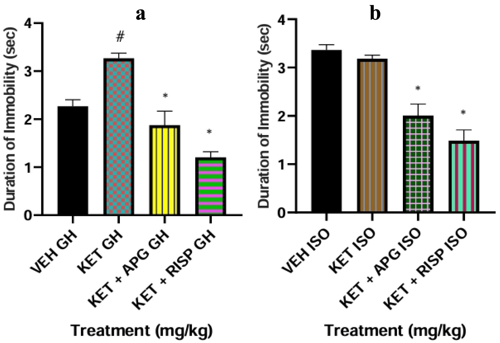 Click for large image | Figure 2. Effect of APG on immobility in weanling rats subjected to two-hit model of neurodevelopmental psychosis in FST. (a) Group-housed animals. (b) Isolated-rearing animals. Bar represents the mean ± SEM of eight animals. *P < 0.05 as compared with KET GH/KET ISO followed by ANOVA and Tukey’s post hoc test for multiple comparisons. #P < 0.05 as compared with VEH GH/VEH ISO followed by ANOVA and Tukey’s post hoc test for multiple comparisons. APG: apigenin; ANOVA: analysis of variance; FST: forced swim test; GH: group-housed; ISO: isolated; KET: ketamine; RISP: risperidone; SEM: standard error of the mean; VEH: vehicle. |
Effect of apigenin on GSH level in amygdala of weanling rats in two-hit model of neurodevelopmental psychosis
Group-housed rats treated with ketamine alone showed a marked reduction in GSH levels compared with vehicle-treated controls (#P < 0.0001; F(3,16) = 1,903). Treatment with apigenin or risperidone significantly increased GSH levels relative to the ketamine-only group (*P < 0.0001). A similar pattern was observed in socially isolated rats, where apigenin and risperidone significantly elevated GSH levels compared with ketamine alone (*P < 0.0001) (Fig. 3).
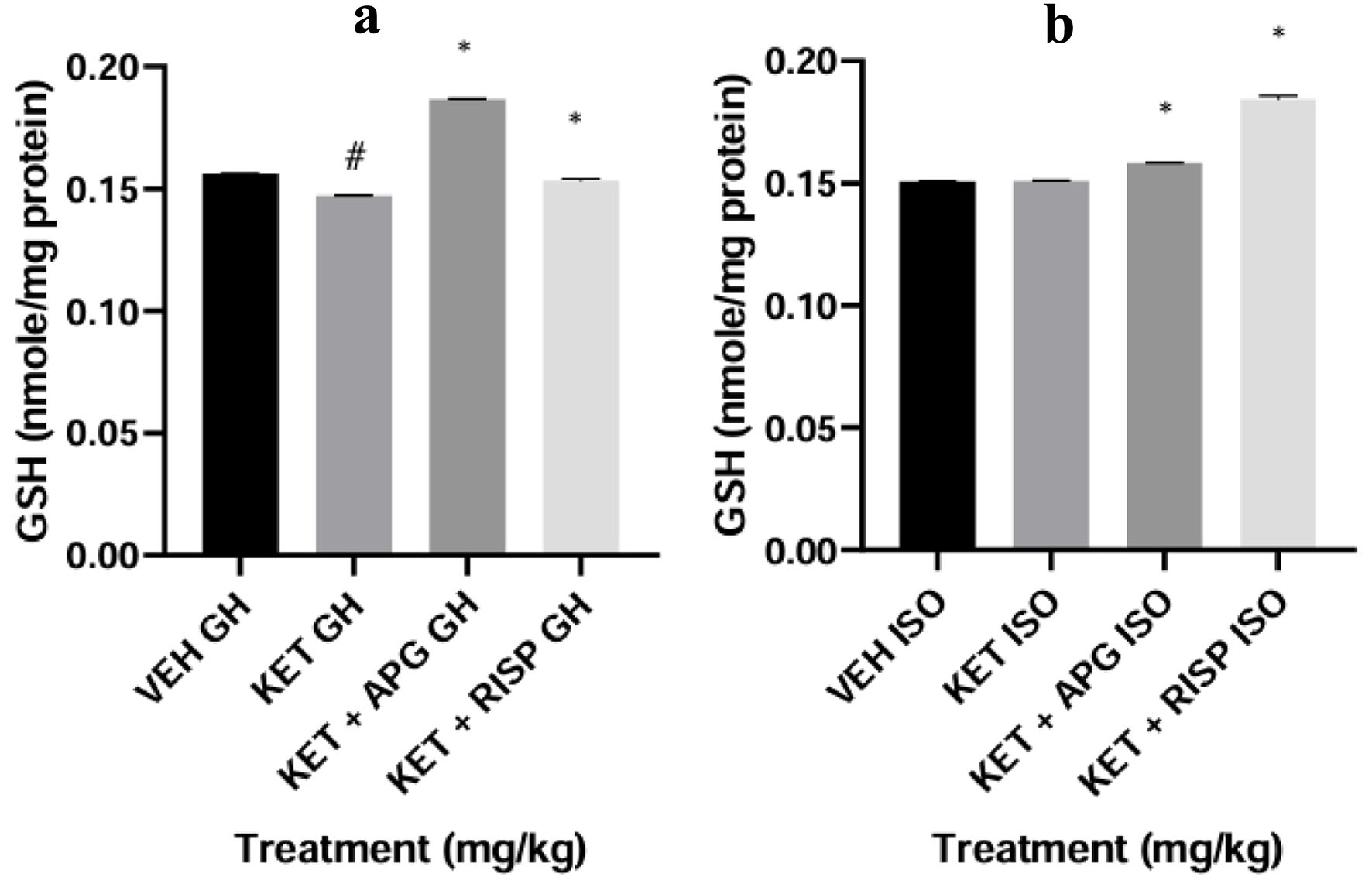 Click for large image | Figure 3. Effect of APG on reduced GSH level in amygdala of weanling rats in two-hit model of neurodevelopmental psychosis. (a) Group-housed animals. (b) Isolated-rearing animals. Bar represents the mean ± SEM of eight animals. *P < 0.05 as compared with KET GH/KET ISO followed by ANOVA and Tukey’s post hoc test for multiple comparisons. #P < 0.05 as compared with VEH GH/VEH ISO followed by ANOVA and Tukey’s post hoc test for multiple comparisons. APG: apigenin; ANOVA: analysis of variance; GH: group-housed; GSH: reduced glutathione; ISO: isolated; KET: ketamine; RISP: risperidone; SEM: standard error of the mean; VEH: vehicle. |
Effect of apigenin on GSH level in hippocampus of weanling rats in two-hit model of neurodevelopmental psychosis
Ketamine significantly reduced GSH levels in group-housed rats compared with vehicle controls (F(3,16) = 5,752, #P < 0.0001). This effect was significantly reversed by apigenin and risperidone (*P < 0.0001). Likewise, in socially isolated rats, both treatments significantly increased hippocampal GSH levels relative to ketamine-treated animals (*P < 0.0001) (Fig. 4).
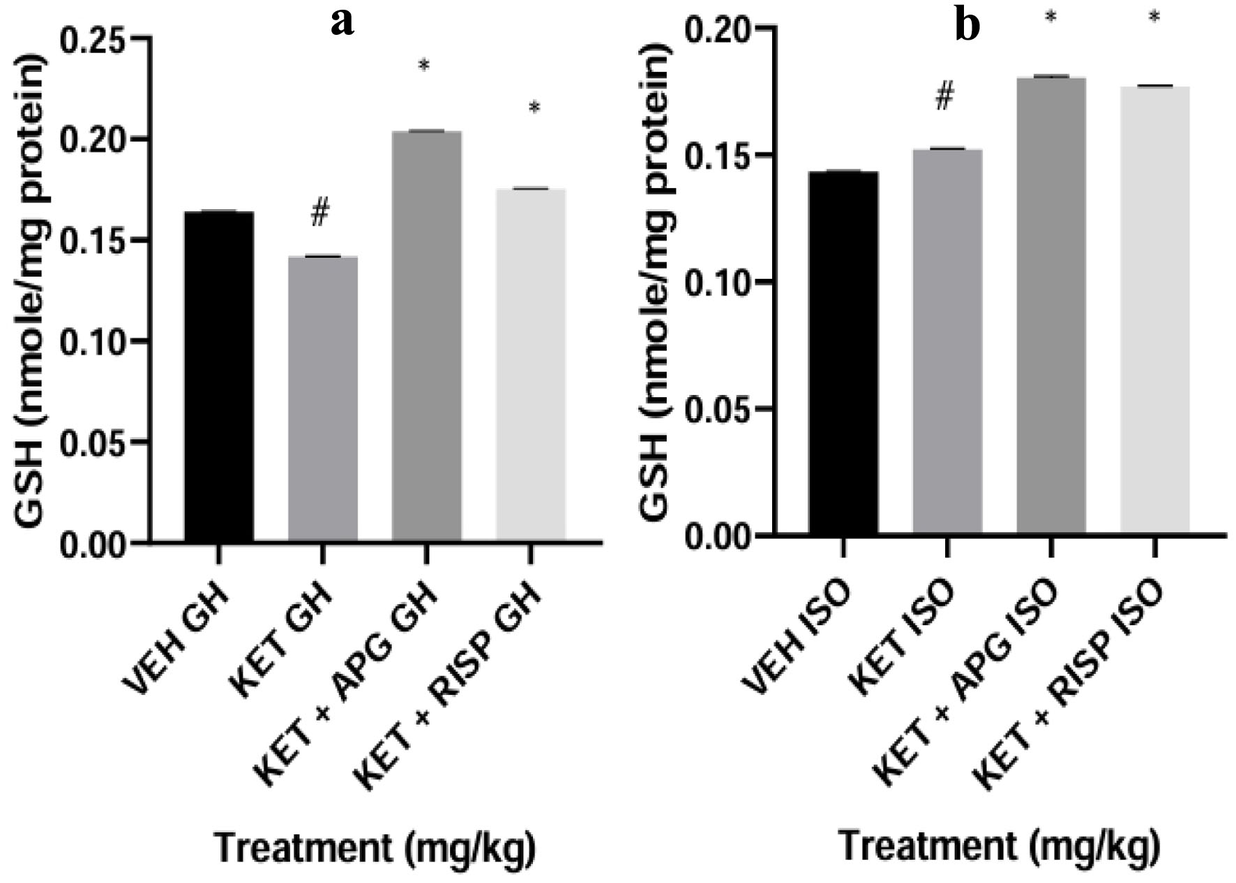 Click for large image | Figure 4. Effect of APG on GSH level in hippocampus of weanling rats subjected to two-hit model of neurodevelopmental psychosis. (a) Group-housed animals. (b) Isolated-rearing animals. Bar represents the mean ± SEM of eight animals. *P < 0.05 as compared with KET GH/KET ISO followed by ANOVA and Tukey’s post hoc test for multiple comparisons. #P < 0.05 as compared with VEH GH/VEH ISO followed by ANOVA and Tukey’s post hoc test for multiple comparisons. APG: apigenin; ANOVA: analysis of variance; GH: group-housed; GSH: reduced glutathione; ISO: isolated; KET: ketamine; RISP: risperidone; SEM: standard error of the mean; VEH: vehicle. |
Effect of apigenin on GSH level in PFC of weanling rats in two-hit model of neurodevelopmental psychosis
Ketamine significantly decreased GSH levels in group-housed rats compared with vehicle-treated controls (F(3,16) = 1,740, #P < 0.0001), while apigenin and risperidone markedly restored GSH levels (*P < 0.0001). In isolated rearing rats, ketamine also significantly altered GSH levels compared with vehicle-treated isolated controls, while apigenin and risperidone significantly increased GSH concentrations relative to the ketamine-only group (*P < 0.0001). These results are presented in Figure 5.
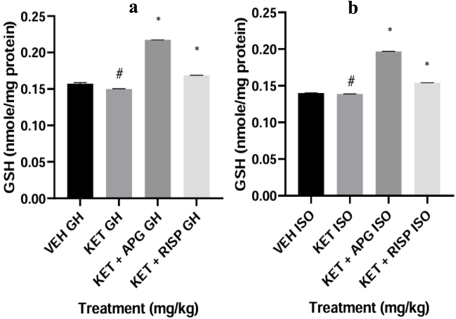 Click for large image | Figure 5. Effect of APG on GSH level in Prefrontal cortex of weanling rats subjected to two-hit model of neurodevelopmental psychosis. (a) Group-housed animals. (b) Isolated-rearing animals. Bar represents the mean ± SEM of eight animals. *P < 0.05 as compared with KET GH/KET ISO followed by ANOVA and Tukey’s post hoc test for multiple comparisons. #P < 0.05 as compared with VEH GH/VEH ISO followed by ANOVA and Tukey’s post hoc test for multiple comparisons. APG: apigenin; ANOVA: analysis of variance; GH: group-housed; GSH: reduced glutathione; ISO: isolated; KET: ketamine; RISP: risperidone; SEM: standard error of the mean; VEH: vehicle. |
Effect of apigenin on MDA level in amygdala of weanling rats in two-hit model of neurodevelopmental psychosis
The levels of MDA were increased in ketamine-treated group-housed rats when compared with vehicle-treated group-housed rats (#P < 0.0001, F(3,16) = 1,029). However, apigenin and risperidone-treated rats showed a decreased level of MDA when compared with ketamine-treated group-housed rats (*P < 0.0001). Furthermore, the ketamine-treated isolated grouped rats showed an increased level of MDA compared with the vehicle-treated isolated-rearing rats (#P < 0.0001, F(3,16) = 2,555). However, apigenin and risperidone-treated isolated grouped rats showed a decreased level of MDA compared with ketamine-treated isolated rats (*P < 0.0001), as depicted in Figure 6.
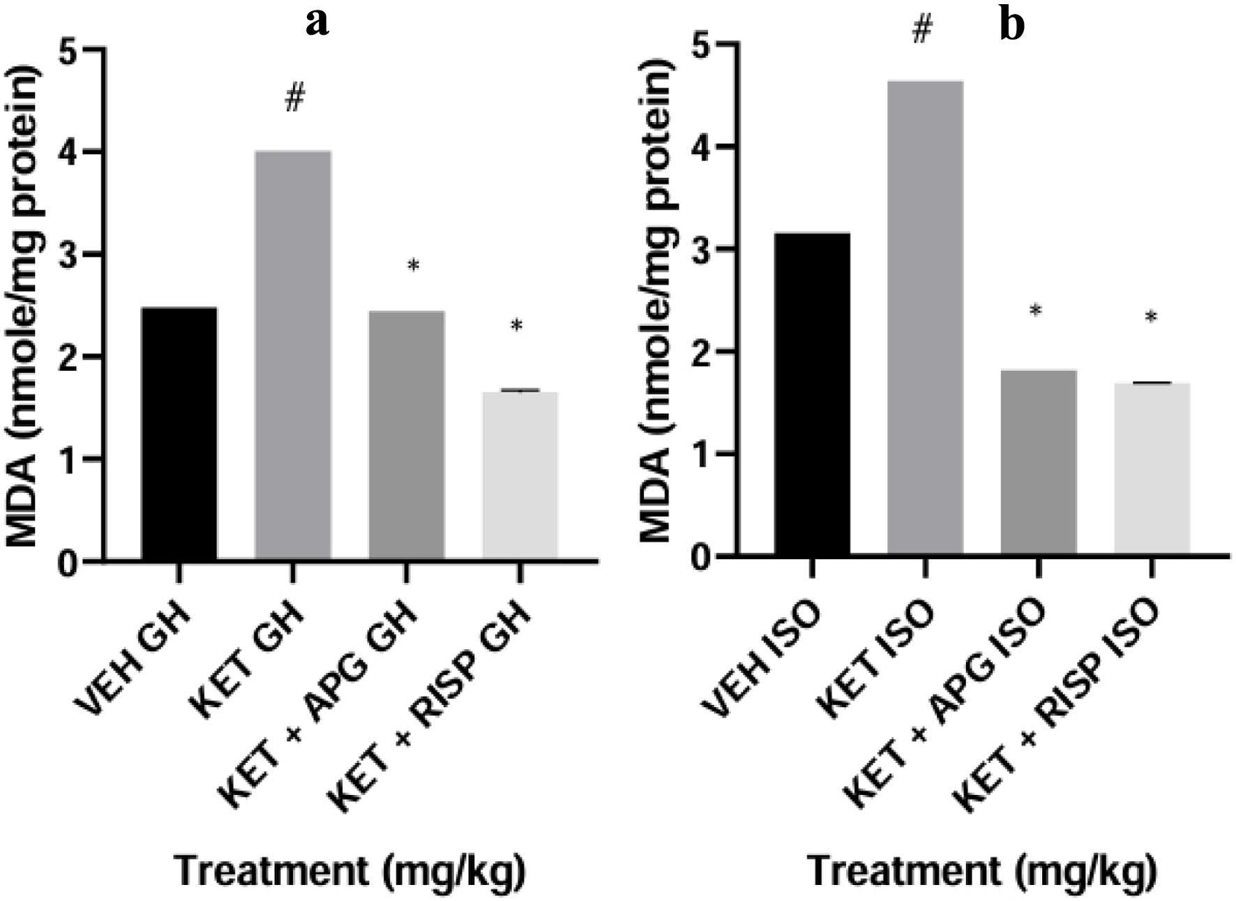 Click for large image | Figure 6. Effect of APG on MDA level in amygdala of weanling rats subjected to two-hit model of neurodevelopmental psychosis. (a) Group-housed animals. (b) Isolated-rearing animals. Bar represents the mean ± SEM of eight animals. *P < 0.05 as compared with KET GH/KET ISO followed by ANOVA and Tukey’s post hoc test for multiple comparisons. #P < 0.05 as compared with VEH GH/VEH ISO followed by ANOVA and Tukey’s post hoc test for multiple comparisons. APG: apigenin; ANOVA: analysis of variance; GH: group-housed; ISO: isolated; KET: ketamine; MDA: malondialdehyde; RISP: risperidone; SEM: standard error of the mean; VEH: vehicle. |
Effect of apigenin on MDA level in hippocampus of weanling rats in two-hit model of neurodevelopmental psychosis
The levels of MDA were increased in ketamine-treated group-housed rats compared with the vehicle-treated group-housed rats (#P < 0.0001, F(3,16) = 2,010). The intervention of apigenin and risperidone reversed the increase in the levels of MDA when compared with ketamine-treated group-housed rats (*P < 0.0001). Furthermore, there was an increase in the level of MDA in ketamine-isolated group-housed rats when compared with the vehicle-treated isolated-rearing rats (#P < 0.0001, F(3,16) = 8,776); however, the intervention by apigenin and risperidone-treated isolated grouped rats showed a decreased MDA level when compared with ketamine-treated isolated rats (*P < 0.0001), as depicted in Figure 7.
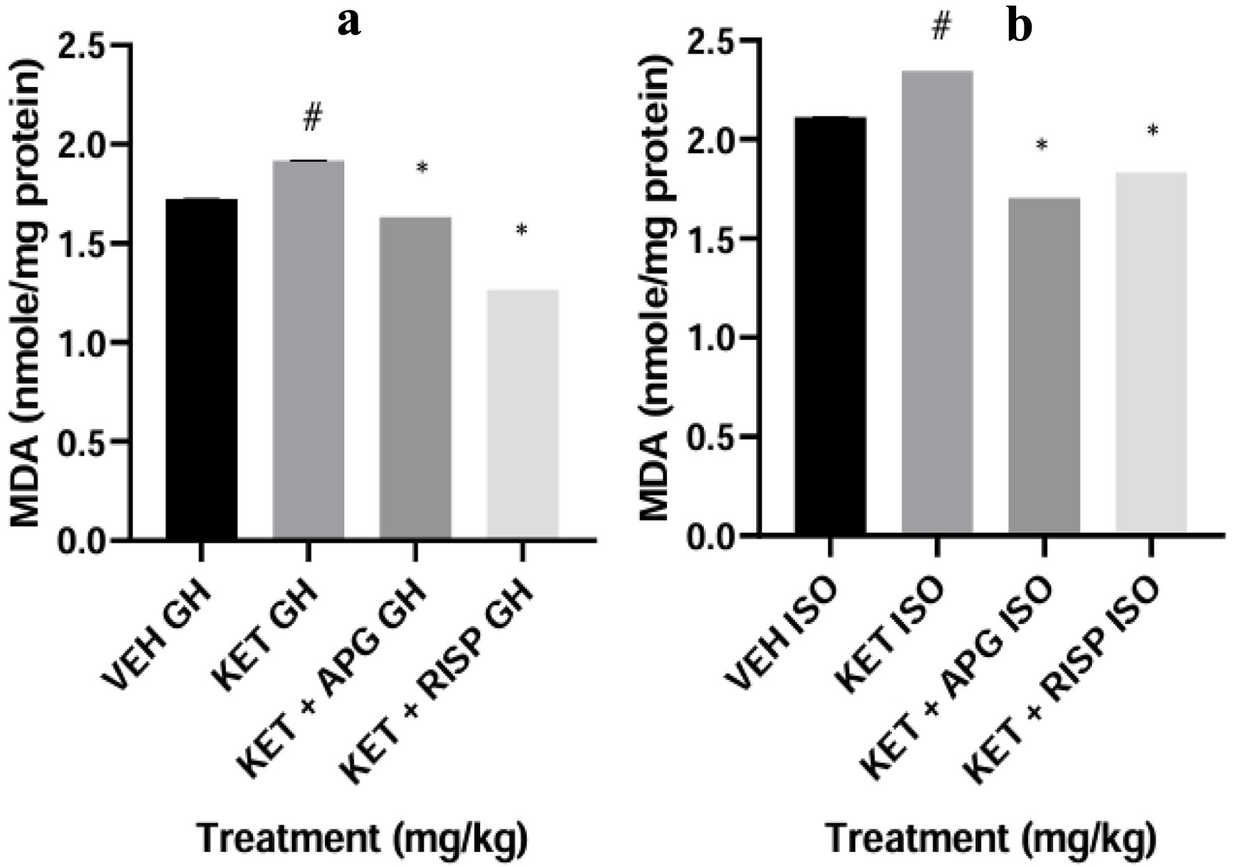 Click for large image | Figure 7. Effect of APG on MDA level in hippocampus of weanling rats subjected to two-hit model of neurodevelopmental psychosis. (a) Group-housed animals. (b) Isolated-rearing animals. Bar represents the mean ± SEM of eight animals. *P < 0.05 as compared with KET GH/KET ISO followed by ANOVA and Tukey’s post hoc test for multiple comparisons. #P < 0.05 as compared with VEH GH/VEH ISO followed by ANOVA and Tukey’s post hoc test for multiple comparisons. APG: apigenin; ANOVA: analysis of variance; GH: group-housed; ISO: isolated; KET: ketamine; MDA: malondialdehyde; RISP: risperidone; SEM: standard error of the mean; VEH: vehicle. |
Effect of apigenin on MDA level in PFC of weanling rats in two-hit model of neurodevelopmental psychosis
The levels of MDA in ketamine-treated group-housed rats were increased when compared with vehicle-treated group-housed rats (#P < 0.0001, F(3,16) = 4,713). The apigenin and risperidone-treated rats showed a decreased MDA level when compared with the ketamine-treated group-housed rats (*P < 0.0001). Furthermore, the levels of MDA were increased in ketamine-isolated group-housed rats when compared with the vehicle-treated isolated rearing (#P < 0.0001, F(3,16) = 1,692); however, the apigenin and risperidone-treated isolated grouped rats showed a decreased level of MDA when compared with ketamine-treated isolated rats (*P < 0.0001), as depicted in Figure 8.
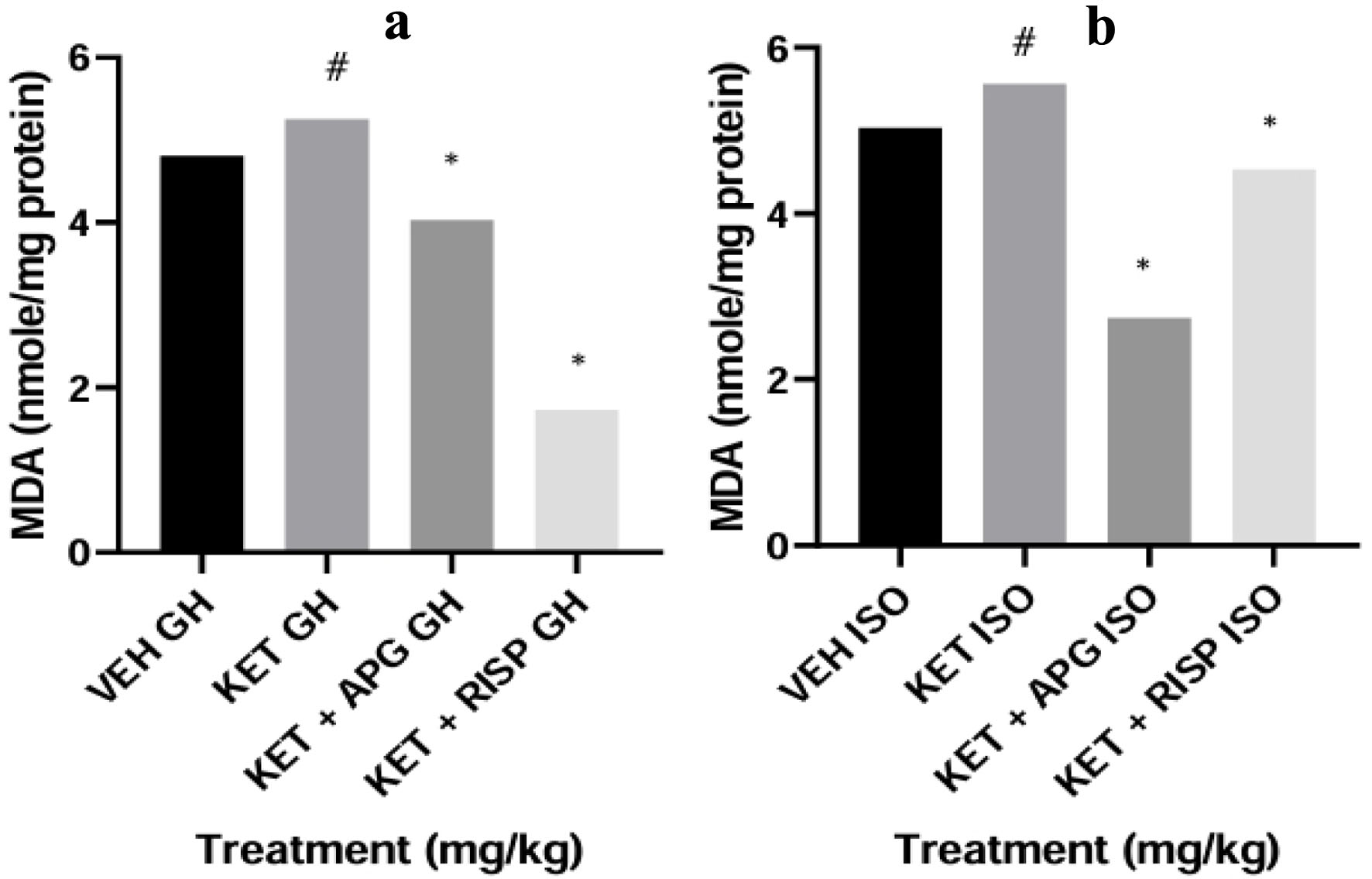 Click for large image | Figure 8. Effect of APG on MDA level in prefrontal cortex of weanling rats subjected to two-hit model of neurodevelopmental psychosis. (a) Group-housed animals. (b) Isolated-rearing animals. Bar represents the mean ± SEM of eight animals. *P < 0.05 as compared with KET GH/KET ISO followed by ANOVA and Tukey’s post hoc test for multiple comparisons. #P < 0.05 as compared with VEH GH/VEH ISO followed by ANOVA and Tukey’s post hoc test for multiple comparisons. APG: apigenin; ANOVA: analysis of variance; GH: group-housed; ISO: isolated; KET: ketamine; MDA: malondialdehyde; RISP: risperidone; SEM: standard error of the mean; VEH: vehicle. |
Effect of apigenin on SOD level in amygdala of weanling rats in two-hit model of neurodevelopmental psychosis
The level of SOD was decreased in ketamine-treated group-housed rats when compared with the vehicle-treated group-housed rats (P < 0.0001, F(3,16) = 1,178). The intervention by apigenin and risperidone-treated rats showed an increased level of SOD when compared with the ketamine-treated group-housed rats (*P < 0.0001). Furthermore, there was no difference between the level of SOD in ketamine-treated isolated grouped rats when compared with vehicle-treated isolated rats; however, apigenin and risperidone-treated isolated grouped rats showed an increased SOD level when compared with the ketamine-treated isolated rats (F(3,16) = 1,692, *P < 0.0001), as depicted in Figure 9.
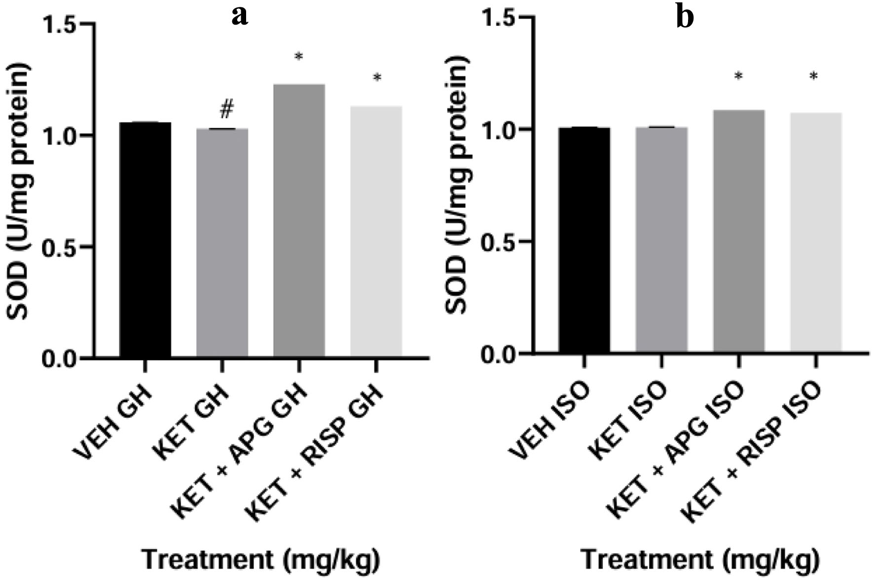 Click for large image | Figure 9. Effect of APG on SOD level in amygdala of weanling rats subjected to two-hit model of neurodevelopmental psychosis. (a) Group-housed animals. (b) Isolated-rearing animals. Bar represents the mean ± SEM of eight animals. *P < 0.05 as compared with KET GH/KET ISO followed by ANOVA and Tukey’s post hoc test for multiple comparisons. #P < 0.05 as compared with VEH GH/VEH ISO followed by ANOVA and Tukey’s post hoc test for multiple comparisons. APG: apigenin; ANOVA: analysis of variance; GH: group-housed; ISO: isolated; KET: ketamine; RISP: risperidone; SEM: standard error of the mean; SOD: superoxide dismutase; VEH: vehicle. |
Effect of apigenin on SOD level in hippocampus of weanling rats in two-hit model of neurodevelopmental psychosis
There was no difference in the level of SOD in the hippocampus of ketamine-treated group-housed rats compared with the vehicle-treated group-housed rats; however, theapigenin and risperidone-treated rats showed an increased level of SOD when compared with ketamine-treated group-housed rats (F(3,16) = 2,531, *P < 0.0001). Similarly, there was no difference in the level of SOD in the hippocampus of ketamine-isolated group-housed rats when compared with the vehicle-treated isolated rearing rats; however, apigenin and risperidone-treated isolated grouped rats showed an increased level of SOD when compared with ketamine-treated isolated rats (F(3,16) = 554.8, *P < 0.0001), as depicted in Figure 10.
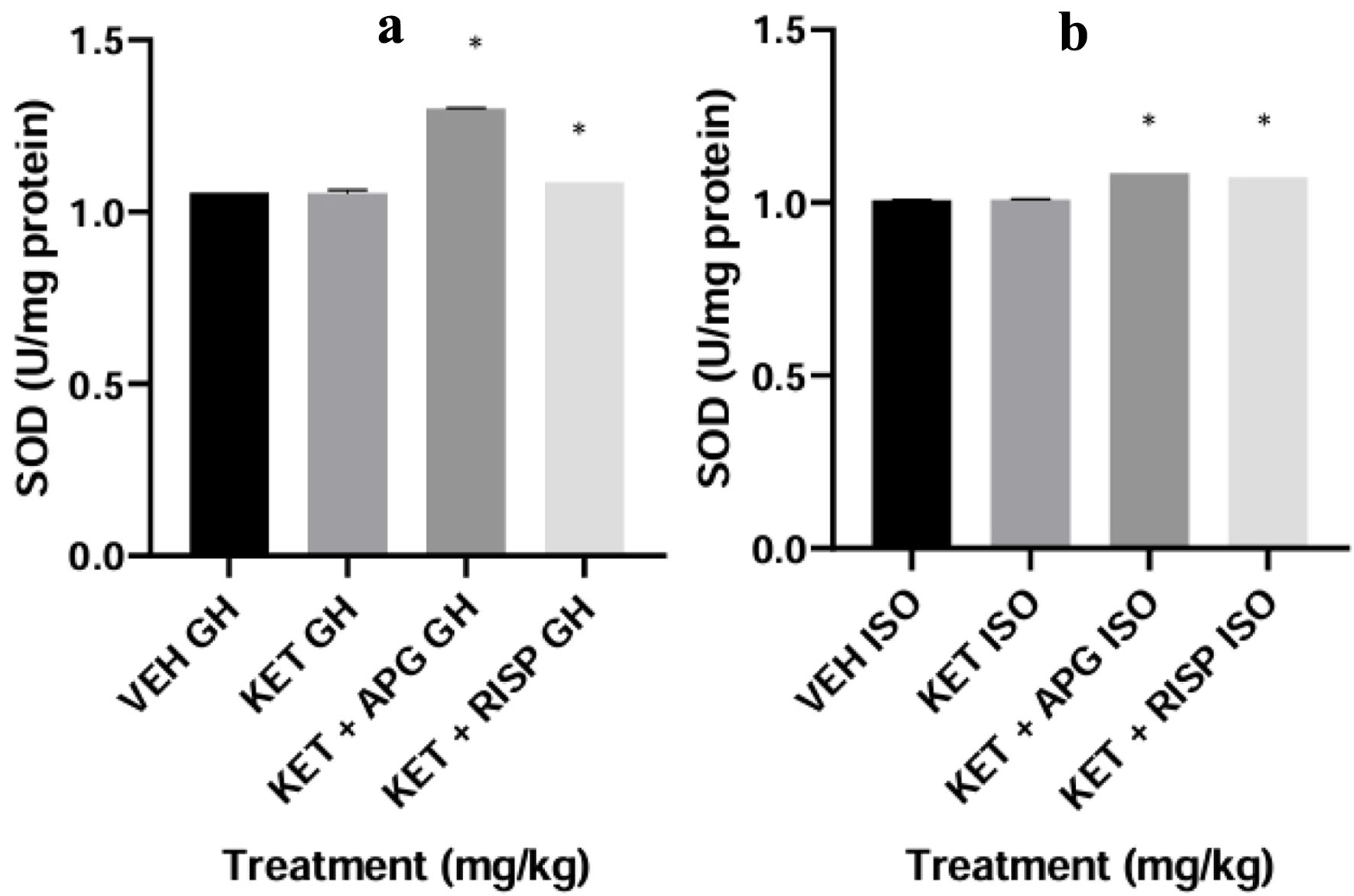 Click for large image | Figure 10. Effect of APG on SOD level in hippocampus of weanling rats subjected to two-hit model of neurodevelopmental psychosis. (a) Group-housed animals. (b) Isolated-rearing animals. Bar represents the mean ± SEM of eight animals. *P < 0.05 as compared with KET GH/KET ISO followed by ANOVA and Tukey’s post hoc test for multiple comparisons. #P < 0.05 as compared with VEH GH/VEH ISO followed by ANOVA and Tukey’s post hoc test for multiple comparisons. APG: apigenin; ANOVA: analysis of variance; GH: group-housed; ISO: isolated; KET: ketamine; RISP: risperidone; SEM: standard error of the mean; SOD: superoxide dismutase; VEH: vehicle. |
Effect of apigenin on SOD level in PFC of weanling rats in two-hit model of neurodevelopmental psychosis
The level of SOD was decreased in the PFC of ketamine-treated group-housed rats when compared with the vehicle-treated group-housed rats (#P < 0.0001, F(3,16) = 4,034). The apigenin and risperidone-treated rats showed an increased level of SOD when compared with ketamine-treated group-housed rats (*P < 0.0001). Similarly, there was a decreased level of SOD in ketamine-isolated group-housed rats when compared with the vehicle-treated isolated-rearing rats (#P< 0.001, F(3,16) = 7,862). However, apigenin and risperidone-treated isolated grouped rats showed an increased level of SOD when compared with ketamine-treated isolated rats (*P < 0.0001), as depicted in Figure 11.
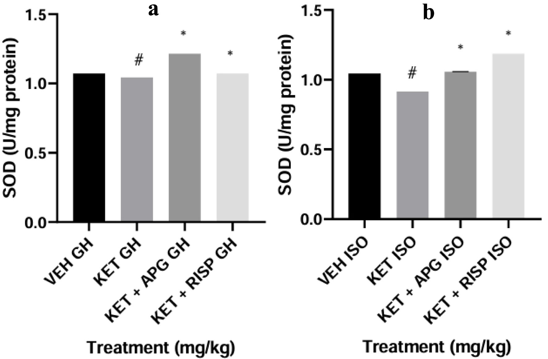 Click for large image | Figure 11. Effect of APG on SOD level in Prefrontal cortex of weanling rats subjected to two-hit model of neurodevelopmental psychosis. (a) Group-housed animals. (b) Isolated-rearing animals. Bar represents the mean ± SEM of eight animals. *P < 0.05 as compared with KET GH/KET ISO followed by ANOVA and Tukey’s post hoc test for multiple comparisons. #P < 0.05 as compared with VEH GH/VEH ISO followed by ANOVA and Tukey’s post hoc test for multiple comparisons. APG: apigenin; ANOVA: analysis of variance; GH: group-housed; ISO: isolated; KET: ketamine; RISP: risperidone; SEM: standard error of the mean; SOD: superoxide dismutase; VEH: vehicle. |
Effect of apigenin on NO level in amygdala of weanling rats in two-hit model of neurodevelopmental psychosis
The level of NO was increased in ketamine-treated group-housed rats compared with the vehicle-treated group-housed rats (#P < 0.0001, F(3,16) = 4,772). The apigenin and risperidone-treated rats showed a decreased NO level when compared with ketamine-treated group-housed rats (*P < 0.0001). Similarly, the level of NO was increased in ketamine-isolated group-housed rats when compared with the vehicle-treated isolated-rearing rats (#P< 0.001, F(3,16) = 1,131). However, the apigenin and risperidone-treated isolated grouped rats showed a decreased level of NO when compared with ketamine-treated isolated rats (*P < 0.0001), as depicted in Figure 12.
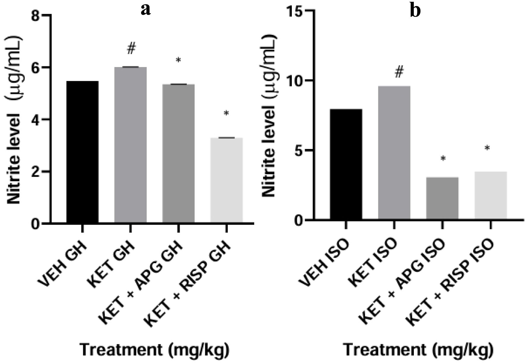 Click for large image | Figure 12. Effect of APG on nitrite level in amygdala of weanling rats subjected to two-hit model of neurodevelopmental psychosis. (a) Group-housed animals. (b) Isolated-rearing animals. Bar represents the mean ± SEM of eight animals. *P < 0.05 as compared with KET GH/KET ISO followed by ANOVA and Tukey’s post hoc test for multiple comparisons. #P < 0.05 as compared with VEH GH/VEH ISO followed by ANOVA and Tukey’s post hoc test for multiple comparisons. APG: apigenin; ANOVA: analysis of variance; GH: group-housed; ISO: isolated; KET: ketamine; RISP: risperidone; SEM: standard error of the mean; VEH: vehicle. |
Effect of apigenin on NO level in hippocampus of weanling rats in two-hit model of neurodevelopmental psychosis
The level of NO was increased in ketamine-treated group-housed rats when compared with the vehicle-treated group-housed rats (#P < 0.0001, F(3,16) = 2,687). The apigenin and risperidone-treated rats showed a decreased NO level when compared with ketamine-treated group-housed rats (*P < 0.0001). Similarly, the level of NO was increased in ketamine-isolated group-housed rats when compared with vehicle-treated isolated-rearing rats (#P< 0.001, F(3,16) = 2,156). However, apigenin and risperidone-treated isolated grouped rats showed a decreased level of NO when compared with ketamine-treated isolated rats (*P < 0.0001), as depicted in Figure 13.
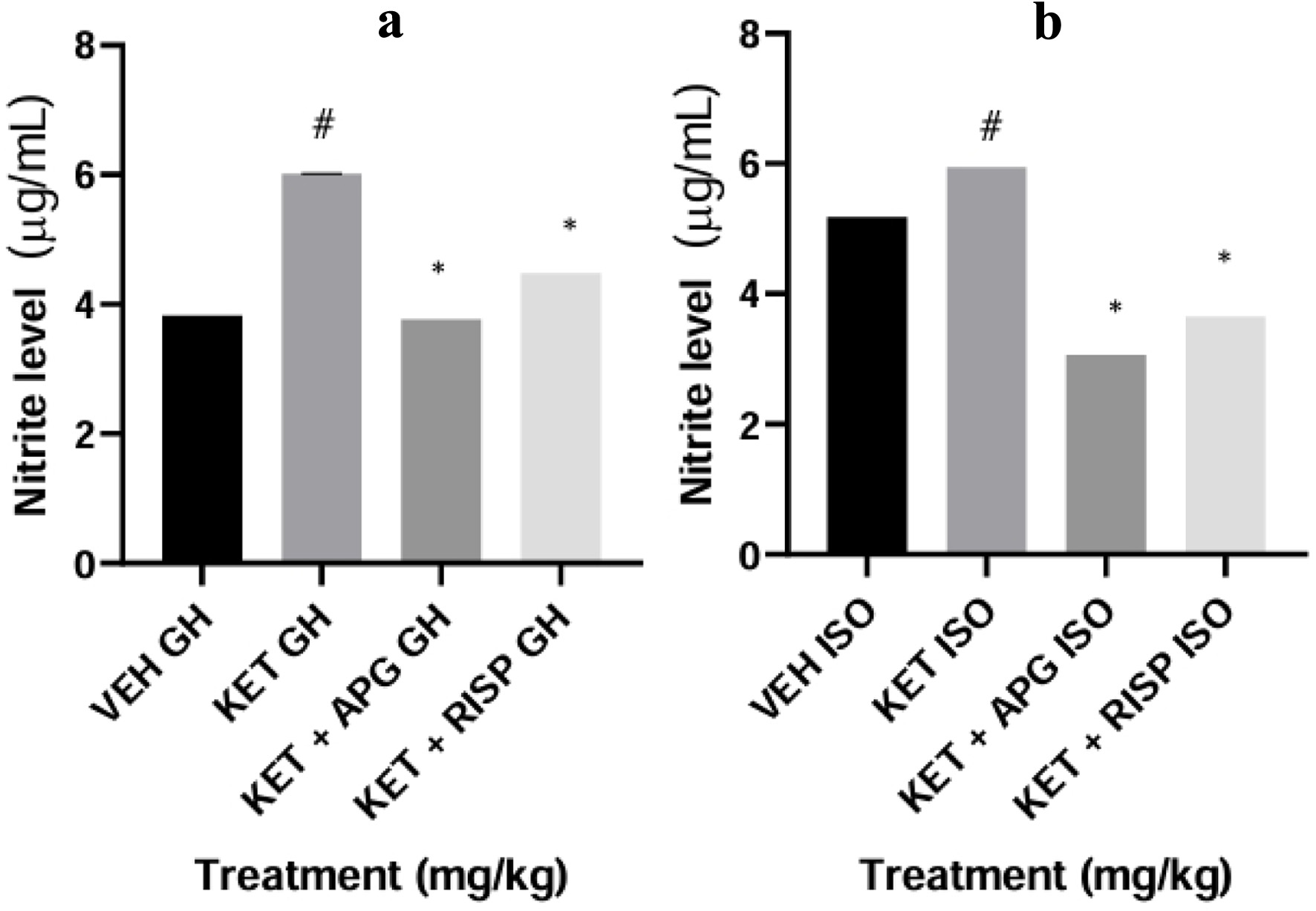 Click for large image | Figure 13. Effect of APG on nitrite level in hippocampus of weanling rats subjected to two-hit model of neurodevelopmental psychosis. (a) Group-housed animals. (b) Isolated-rearing animals. Bar represents the mean ± SEM of eight animals. *P < 0.05 as compared with KET GH/KET ISO followed by ANOVA and Tukey’s post hoc test for multiple comparisons. #P < 0.05 as compared with VEH GH/VEH ISO followed by ANOVA and Tukey’s post hoc test for multiple comparisons. APG: apigenin; ANOVA: analysis of variance; GH: group-housed; ISO: isolated; KET: ketamine; RISP: risperidone; SEM: standard error of the mean; VEH: vehicle. |
Effect of apigenin on NO level in PFC of weanling rats in two-hit model of neurodevelopmental psychosis
The NO level in ketamine-treated group-housed rats was increased compared with the vehicle-treated group-housed rats (#P < 0.0001, F(3,16) = 5,407). The intervention of apigenin and risperidone produced a decrease in the NO level when compared with ketamine-treated group-housed rats (*P < 0.0001). Similarly, the NO level in ketamine-isolated group rats was increased when compared with the vehicle-treated isolated-rearing rats (#P< 0.001, F(3,16) = 3,160). However, the apigenin and risperidone-treated isolated grouped rats showed a decreased NO level when compared with ketamine-treated isolated rats (*P < 0.0001), as depicted in Figure 14.
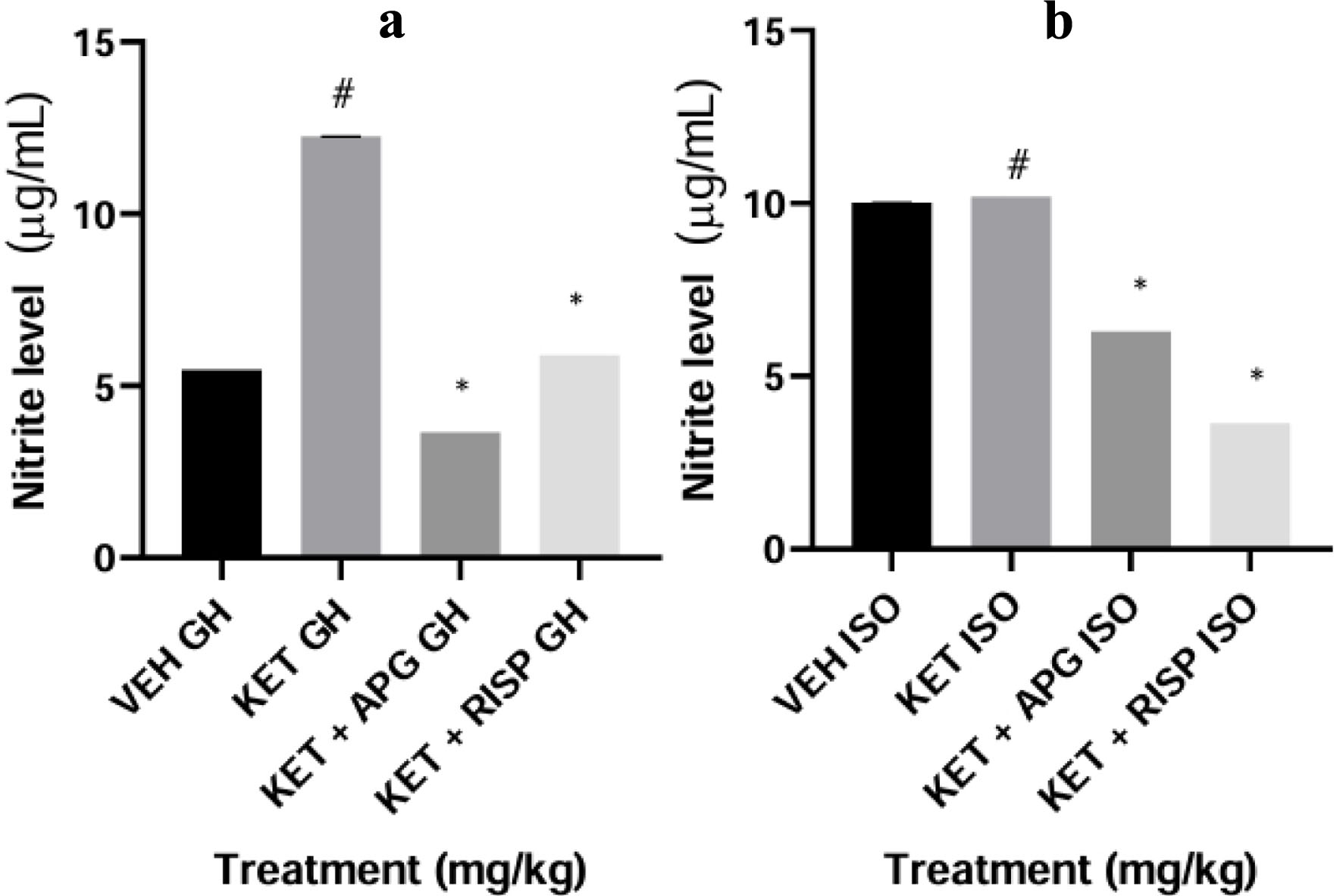 Click for large image | Figure 14. Effect of APG on nitrite level in prefrontal cortex of weanling rats subjected to two-hit model of neurodevelopmental psychosis. (a) Group-housed animals. (b) Isolated-rearing animals. Bar represents the mean ± SEM of eight animals. *P < 0.05 as compared with KET GH/KET ISO followed by ANOVA and Tukey’s post hoc test for multiple comparisons. #P < 0.05 as compared with VEH GH/VEH ISO followed by ANOVA and Tukey’s post hoc test for multiple comparisons. APG: apigenin; ANOVA: analysis of variance; GH: group-housed; ISO: isolated; KET: ketamine; RISP: risperidone; SEM: standard error of the mean; VEH: vehicle. |
Effect of apigenin on AChE activity on amygdala of weanling rats in two-hit model of neurodevelopmental psychosis
The activity of ACHE was increased in ketamine-treated group-housed rats when compared with vehicle-treated group-housed rats (#P < 0.0001, F(3,16) = 680.4). The apigenin and risperidone-treated rats showed decreased activity of AChE compared with ketamine-treated group-housed rats (*P < 0.0001). Conversely, the activity of AChE was lowered in ketamine-isolated group rats when compared with the vehicle-treated isolated-rearing rats (#P< 0.001, F(3,16) = 753.2). Similarly, the apigenin and risperidone-treated isolated grouped rats also showed a decreased activity of AChE (*P < 0.0001), as depicted in Figure 15.
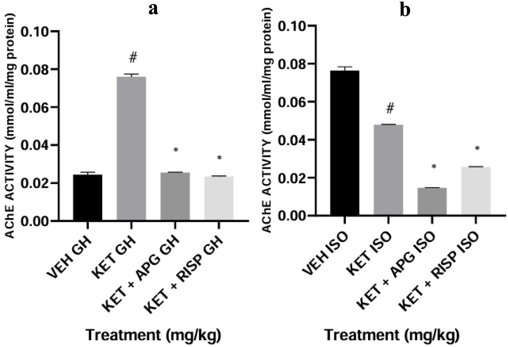 Click for large image | Figure 15. Effect of APG on AChE activity on amygdala of weanling rats subjected to two-hit model of neurodevelopmental psychosis. (a) Group-housed animals. (b) Isolated-rearing animals. Bar represents the mean ± SEM of eight animals. *P < 0.05 as compared with KET GH/KET ISO followed by ANOVA and Tukey’s post hoc test for multiple comparisons. #P < 0.05 as compared with VEH GH/VEH ISO followed by ANOVA and Tukey’s post hoc test for multiple comparisons. AChE: acetylcholinesterase; APG: apigenin; ANOVA: analysis of variance; GH: group-housed; ISO: isolated; KET: ketamine; RISP: risperidone; SEM: standard error of the mean; VEH: vehicle. |
Effect of apigenin on AChE activity on hippocampus of weanling rats in two-hit model of neurodevelopmental psychosis
The activity of AChE was decreased in ketamine-treated group-housed rats when compared with vehicle-treated group-housed rats (#P < 0.0001, F(3,16) = 4,070). Similarly, the apigenin and risperidone-treated rats also showed a decreased AChE activity (*P < 0.0001). Similarly, the activity of AChE was decreased in ketamine-isolated grouped rats when compared with the vehicle-treated isolated-rearing rats (#P< 0.001, F(3,16) = 1,081). Similarly, the apigenin and risperidone-treated isolated grouped rats also showed a decreased activity of AChE (*P < 0.0001), as depicted in Figure 16.
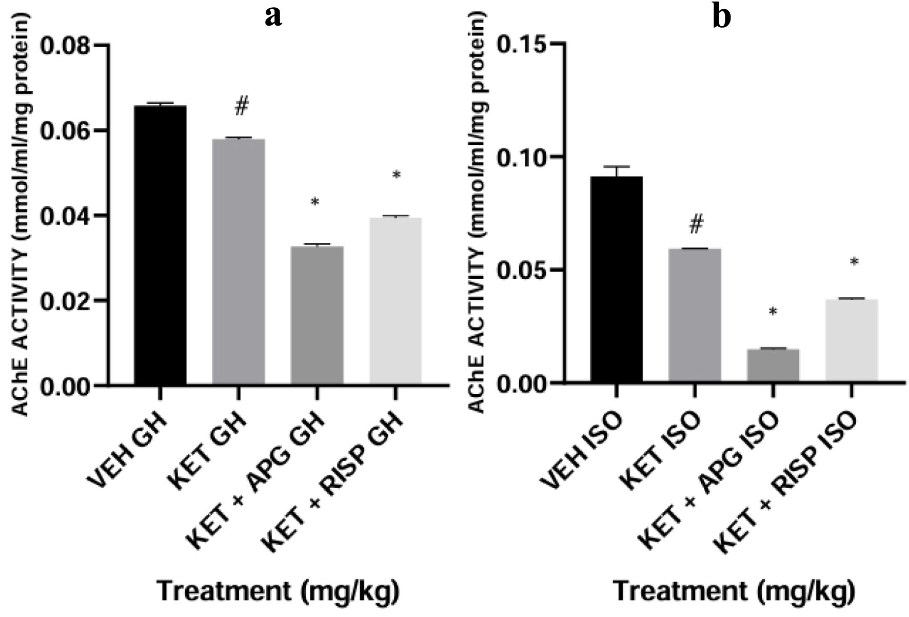 Click for large image | Figure 16. Effect of APG on AChE activity on hippocampus of weanling rats subjected to two-hit model of neurodevelopmental psychosis. (a) Group-housed animals. (b) Isolated-rearing animals. Bar represents the mean ± SEM of eight animals. *P < 0.05 as compared with KET GH/KET ISO followed by ANOVA and Tukey’s post hoc test for multiple comparisons. #P < 0.05 as compared with VEH GH/VEH ISO followed by ANOVA and Tukey’s post hoc test for multiple comparisons. AChE: acetylcholinesterase; APG: apigenin; ANOVA: analysis of variance; GH: group-housed; ISO: isolated; KET: ketamine; RISP: risperidone; SEM: standard error of the mean; VEH: vehicle. |
Effect of apigenin on AChE activity on PFC of weanling rats in two-hit model of neurodevelopmental psychosis
The activity of AChE was increased in ketamine-treated group-housed rats compared with the vehicle-treated group-housed rats (#P < 0.0001, F(3,16) = 1,014). The apigenin and risperidone-treated rats showed a decreased AChE activity when compared with ketamine-treated grouped rats (*P < 0.0001). Conversely, the activity of AChE was decreased in ketamine-isolated grouped rats; similarly, the apigenin and risperidone-treated isolated grouped rats showed a decreased AChE activity (#P < 0.001, F(3,16) = 4,910) (*P < 0.0001), as depicted in Figure 17.
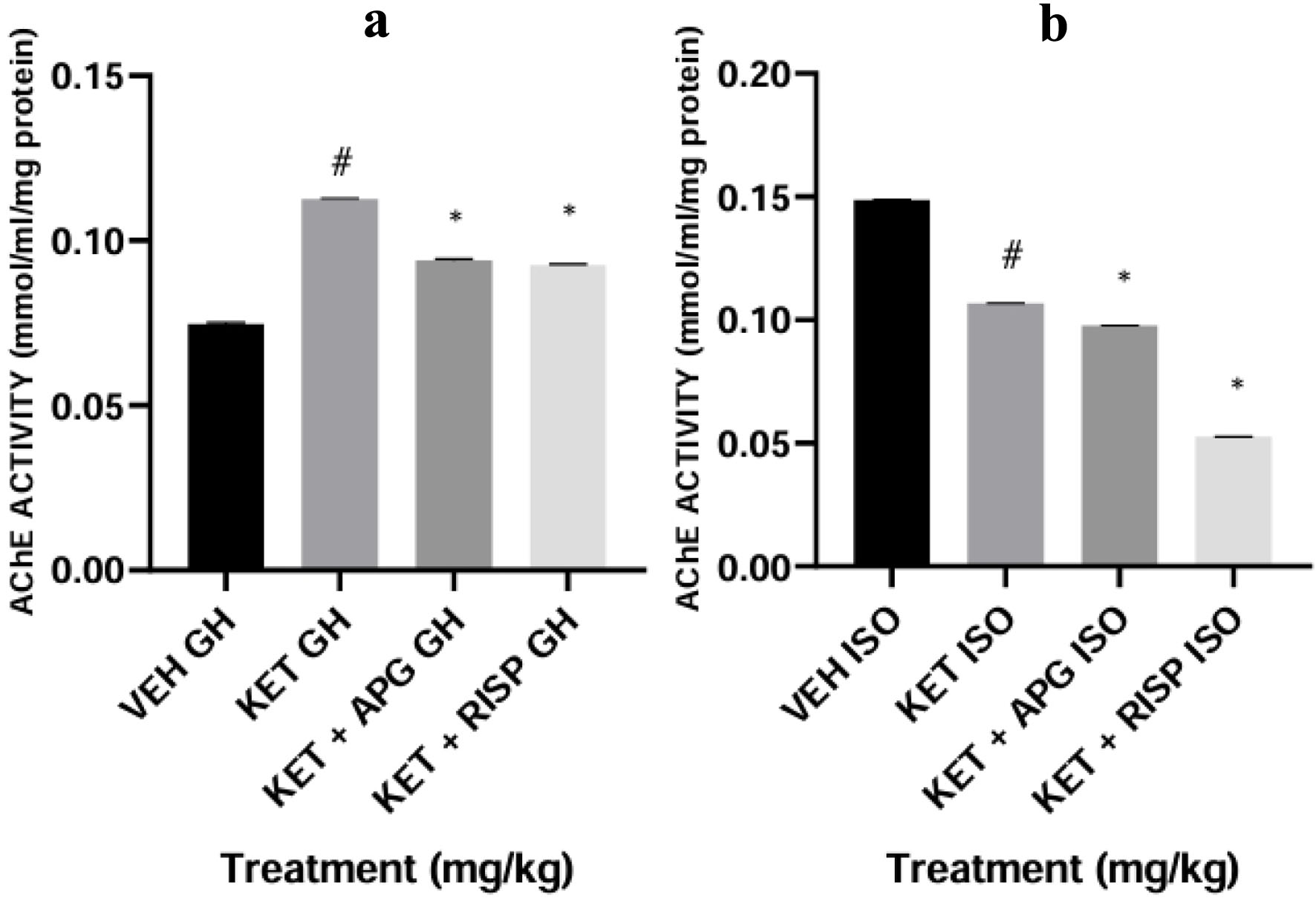 Click for large image | Figure 17. Effect of APG on AChE activity on prefrontal cortex of weanling rats subjected to two-hit model of neurodevelopmental psychosis. (a) Group-housed animals. (b) Isolated-rearing animals. Bar represents the mean ± SEM of eight animals. *P < 0.05 as compared with KET GH/KET ISO followed by ANOVA and Tukey’s post hoc test for multiple comparisons. #P < 0.05 as compared with VEH GH/VEH ISO followed by ANOVA and Tukey’s post hoc test for multiple comparisons. AChE: acetylcholinesterase; APG: apigenin; ANOVA: analysis of variance; GH: group-housed; ISO: isolated; KET: ketamine; RISP: risperidone; SEM: standard error of the mean; VEH: vehicle. |
Effect of apigenin on DA concentration in amygdala of weanling rats in two-hit model of neurodevelopmental psychosis
The DA concentration was decreased in ketamine-treated group-housed rats compared with the vehicle-treated group-housed rats (F(3,16) = 17.72). The apigenin and risperidone-treated rats showed an increased DA concentration compared with the ketamine-treated group-housed (*P < 0.0001). More so, there was no difference in DA concentration in ketamine-isolated grouped rats when compared with the vehicle-treated isolated-rearing rats. However, an increase in DA concentration was shown in apigenin and risperidone-treated isolated grouped rats (*P < 0.0001), as depicted in Figure 18.
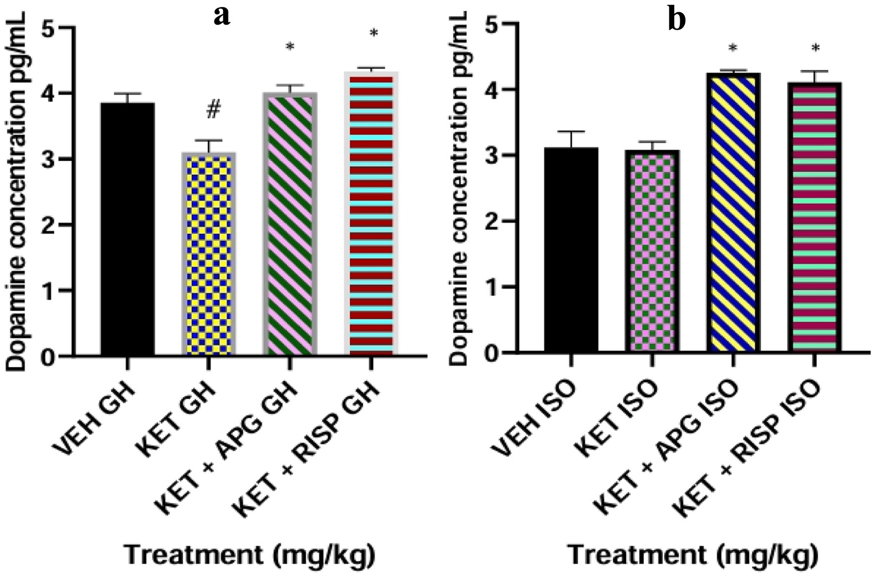 Click for large image | Figure 18. Effect of APG on DA level in Amygdala of weanling rats subjected to two-hit model of neurodevelopmental psychosis. (a) Group-housed animals. (b) Isolated-rearing animals. Bar represents the mean ± SEM of eight animals. *P < 0.05 as compared with KET GH/KET ISO followed by ANOVA and Tukey’s post hoc test for multiple comparisons. #P < 0.05 as compared with VEH GH/VEH ISO followed by ANOVA and Tukey’s post hoc test for multiple comparisons. APG: apigenin; ANOVA: analysis of variance; DA: dopamine; GH: group-housed; ISO: isolated; KET: ketamine; RISP: risperidone; SEM: standard error of the mean; VEH: vehicle. |
Effect of apigenin on DA concentration in hippocampus of weanling rats in two-hit model of neurodevelopmental psychosis
The DA concentration was decreased in ketamine-treated group-housed rats compared with the vehicle-treated group-housed rats (#P < 0.0001, F(3,16) = 142.5). The apigenin and risperidone-treated rats showed an increased DA concentration when compared with ketamine-treated group rats (*P < 0.0001). More so, there was no difference in the DA concentration in ketamine-isolated grouped rats compared with the vehicle-treated isolated-rearing rats. The apigenin and risperidone-treated isolated grouped rats showed an increased DA concentration (*P < 0.0001), as depicted in Figure 19.
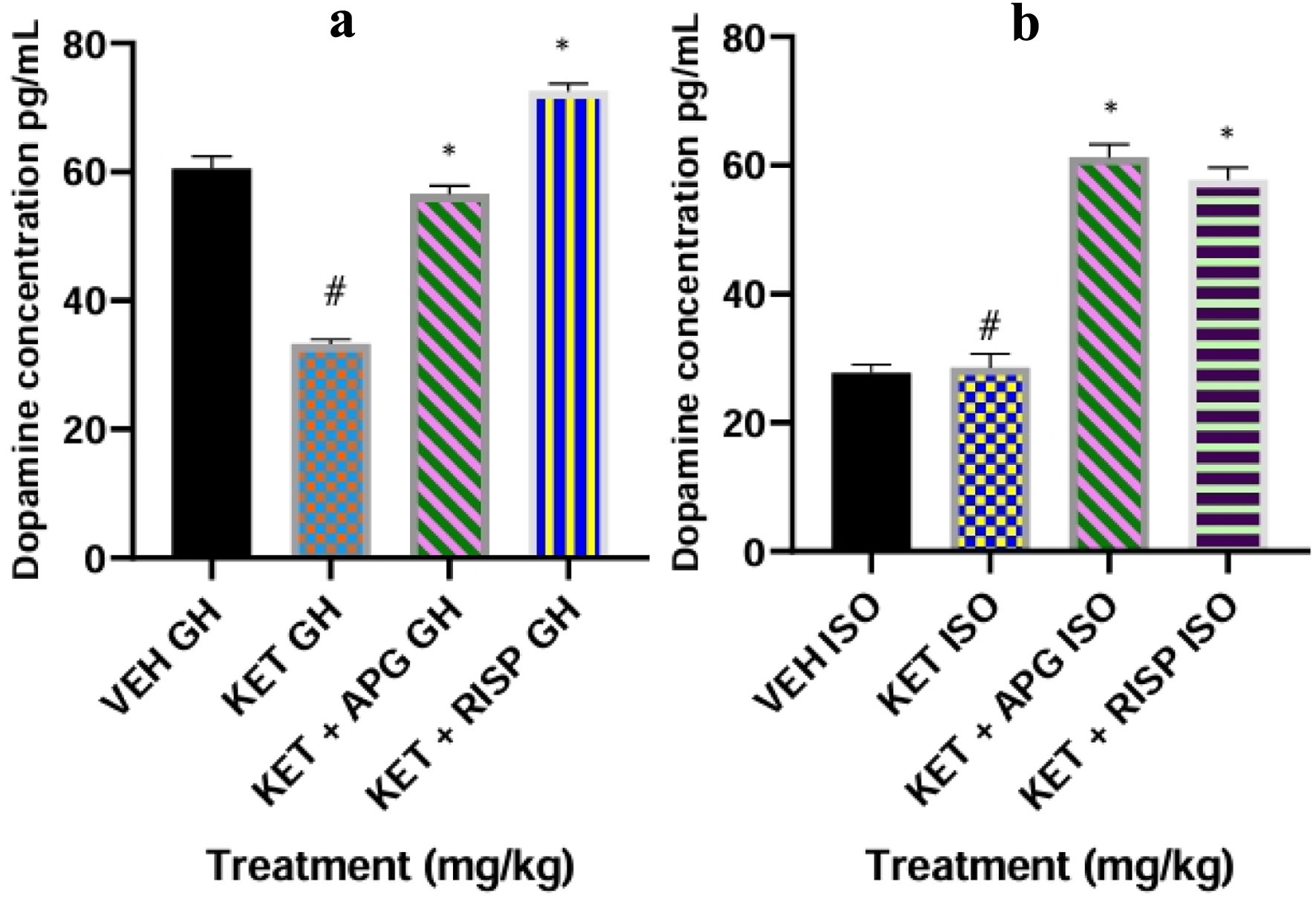 Click for large image | Figure 19. Effect of APG on DA level in Hippocampus of weanling rats subjected to two-hit model of neurodevelopmental psychosis. (a) Group-housed animals. (b) Isolated-rearing animals. Bar represents the mean ± SEM of eight animals. *P < 0.05 as compared with KET GH/KET ISO followed by ANOVA and Tukey’s post hoc test for multiple comparisons. #P < 0.05 as compared with VEH GH/VEH ISO followed by ANOVA and Tukey’s post hoc test for multiple comparisons. APG: apigenin; ANOVA: analysis of variance; DA: dopamine; GH: group-housed; ISO: isolated; KET: ketamine; RISP: risperidone; SEM: standard error of the mean; VEH: vehicle. |
Effect of apigenin on DA concentration in PFC of weanling rats in two-hit model of neurodevelopmental psychosis
The DA concentration was decreased in ketamine-treated group-housed rats when compared with vehicle-treated group-housed rats (#P < 0.0001, F(3,16) = 16.26). The apigenin and risperidone-treated rats showed an increased DA concentration when compared with ketamine-treated grouped rats (*P < 0.0001). More so, there was no difference between the DA concentration in ketamine-isolated grouped rats compared with the vehicle-treated isolated-rearing rats. However, the apigenin and risperidone-treated isolated grouped rats showed an increased DA concentration (*P < 0.0001), as depicted in Figure 20.
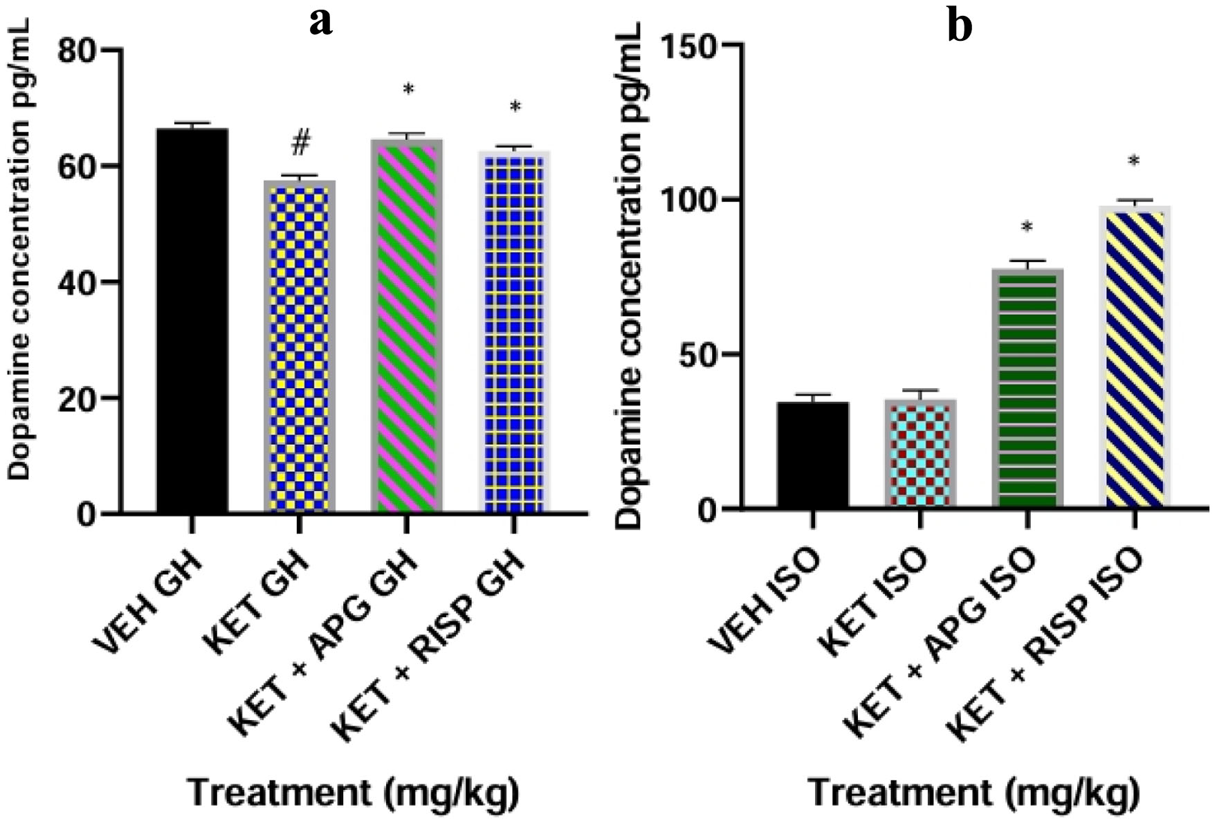 Click for large image | Figure 20. Effect of APG on DA level in prefrontal cortex of weanling rats subjected to two-hit model of neurodevelopmental psychosis. (a) Group-housed animals. (b) Isolated-rearing animals. Bar represents the mean ± SEM of eight animals. *P < 0.05 as compared with KET GH/KET ISO followed by ANOVA and Tukey’s post hoc test for multiple comparisons. #P < 0.05 as compared with VEH GH/VEH ISO followed by ANOVA and Tukey’s post hoc test for multiple comparisons. APG: apigenin; ANOVA: analysis of variance; DA: dopamine; GH: group-housed; ISO: isolated; KET: ketamine; RISP: risperidone; SEM: standard error of the mean; VEH: vehicle. |
Histological assessment (H&E staining) of the amygdala of weanling rats in two-hit model of neurodevelopmental psychosis
Photomicrographs portraying the histological and histomorphological (density of viable neuronal cells) using H&E staining demonstrated that administration of ketamine in group-housed post-weaned rats and isolated-rearing animals significantly (#P < 0.0001) showed cytoarchitectural changes in the amygdala as evidenced in increased population of highly condensed (pyknotic) and angulated neuronal cells in relative to the group that received vehicle. More so, optical densitometry count showed that group that received ketamine in both group-housed animals and isolated-rearing animals showed significant reduction of viable neuronal cell population (*P < 0.0001) compared with apigenin and risperidone-treated group, as depicted in Figures 21 and 22.
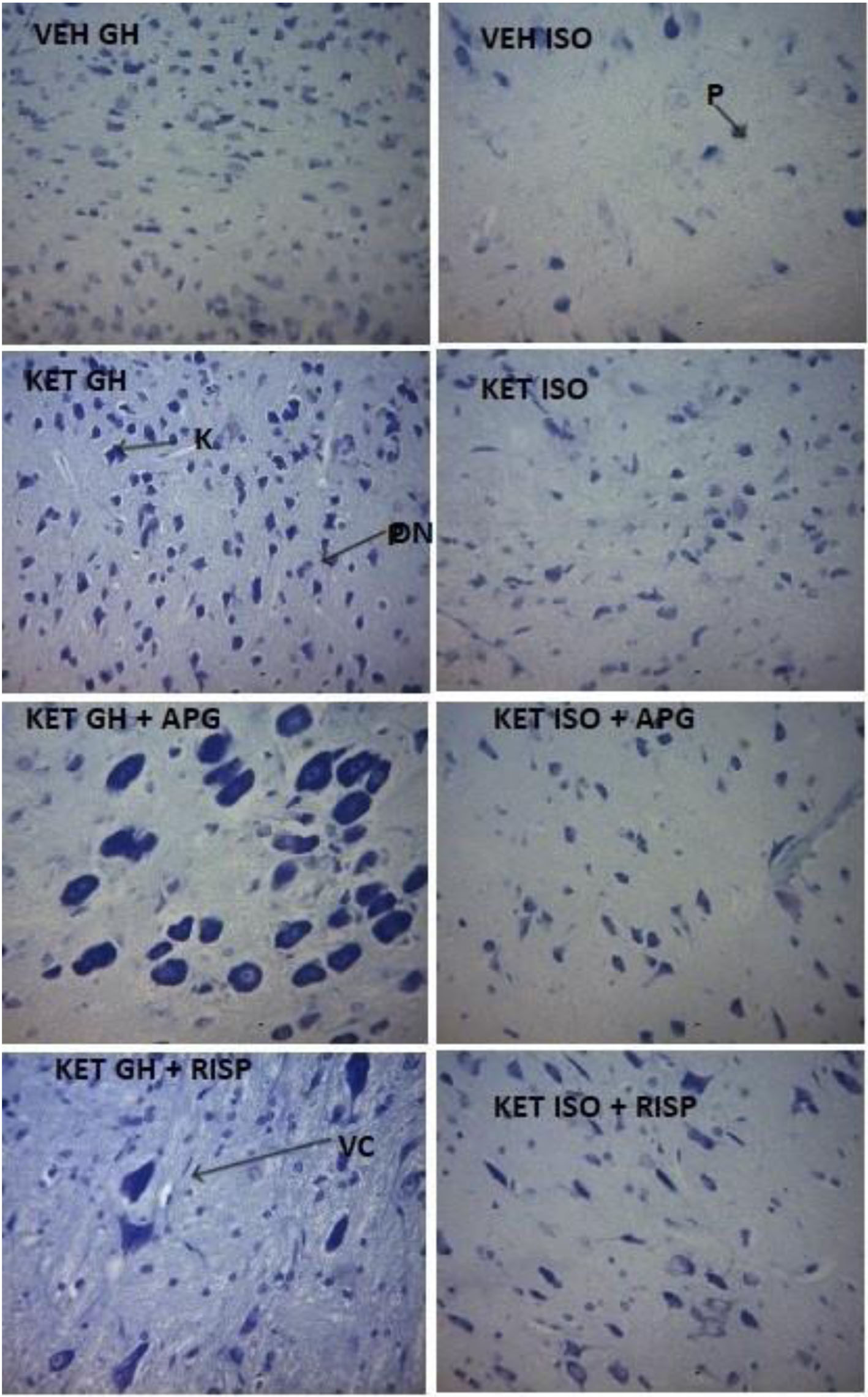 Click for large image | Figure 21. Photomicrographs of amygdala showing surviving neurons in weanling rats subjected to two-hit model of neurodevelopmental psychosis. (a) Group-housed animals. (b) Isolated-rearing animals. APG: apigenin; DN: degenerating neurons; GH: group housed; ISO: isolated; K: karyopyknosis; KET: ketamine; P: pyknosis; RISP: risperidone; VC: vacoulated cytoplasm; VEH: vehicle. |
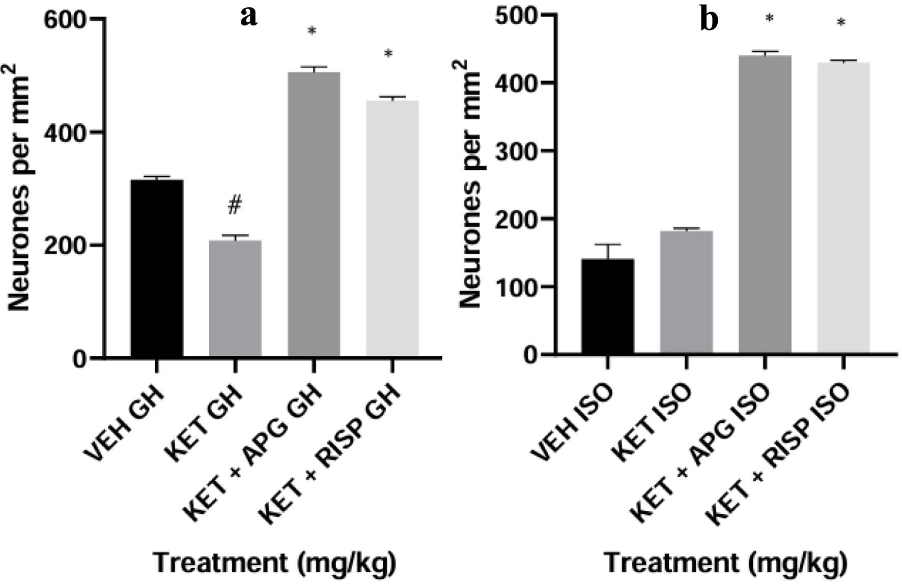 Click for large image | Figure 22. Neuronal density in amygdala of weanling rats subjected to two-hit model of neurodevelopmental psychosis. (a) Group-housed animals. (b) Isolated-rearing animals. Bar represents the mean ± SEM of eight animals. *P < 0.05 as compared with KET GH/KET ISO followed by ANOVA and Tukey’s post hoc test for multiple comparisons. #P < 0.05 as compared with VEH GH/VEH ISO followed by ANOVA and Tukey’s post hoc test for multiple comparisons. APG: apigenin; ANOVA: analysis of variance; GH: group-housed; ISO: isolated; KET: ketamine; RISP: risperidone; SEM: standard error of the mean; VEH: vehicle. |
Histological assessment (H&E staining) of the hippocampus of weanling rats in two-hit model of neurodevelopmental psychosis
Photomicrographs portraying the histological and histomorphological (density of viable neuronal cells) using H&E staining demonstrated that administration of ketamine in group-housed post-weaned rats and isolated-rearing animals significantly (#P < 0.0001) showed cytoarchitectural changes in the hippocampus as evidenced in increased population of highly condensed (pyknotic) and angulated neuronal cells in relative to the group that received vehicle. More so, optical densitometry count showed that group that received ketamine in both group-housed animals and isolated-rearing animals showed significant reduction of viable neuronal cell population (*P < 0.0001) compared with apigenin and risperidone-treated group, as depicted in Figures 23 and 24.
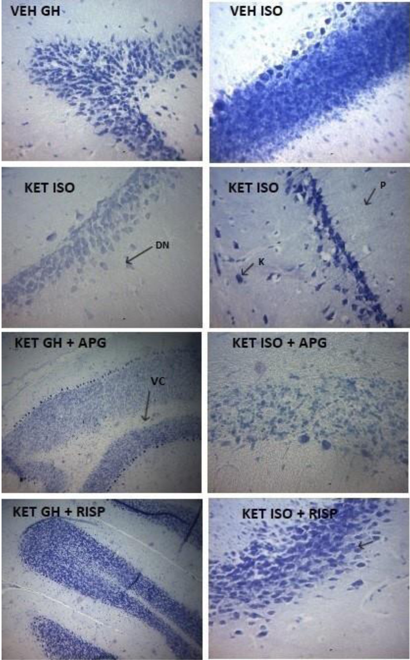 Click for large image | Figure 23. Photomicrographs of hippocampus showing surviving neurons in weanling rats subjected to two-hit model of neurodevelopmental psychosis. APG: apigenin; DN: degenerating neurons; GH: group housed; ISO: isolated; K: karyopyknosis; KET: ketamine; P: pyknosis; RISP: risperidone; VC: vacoulated cytoplasm; VEH: vehicle. |
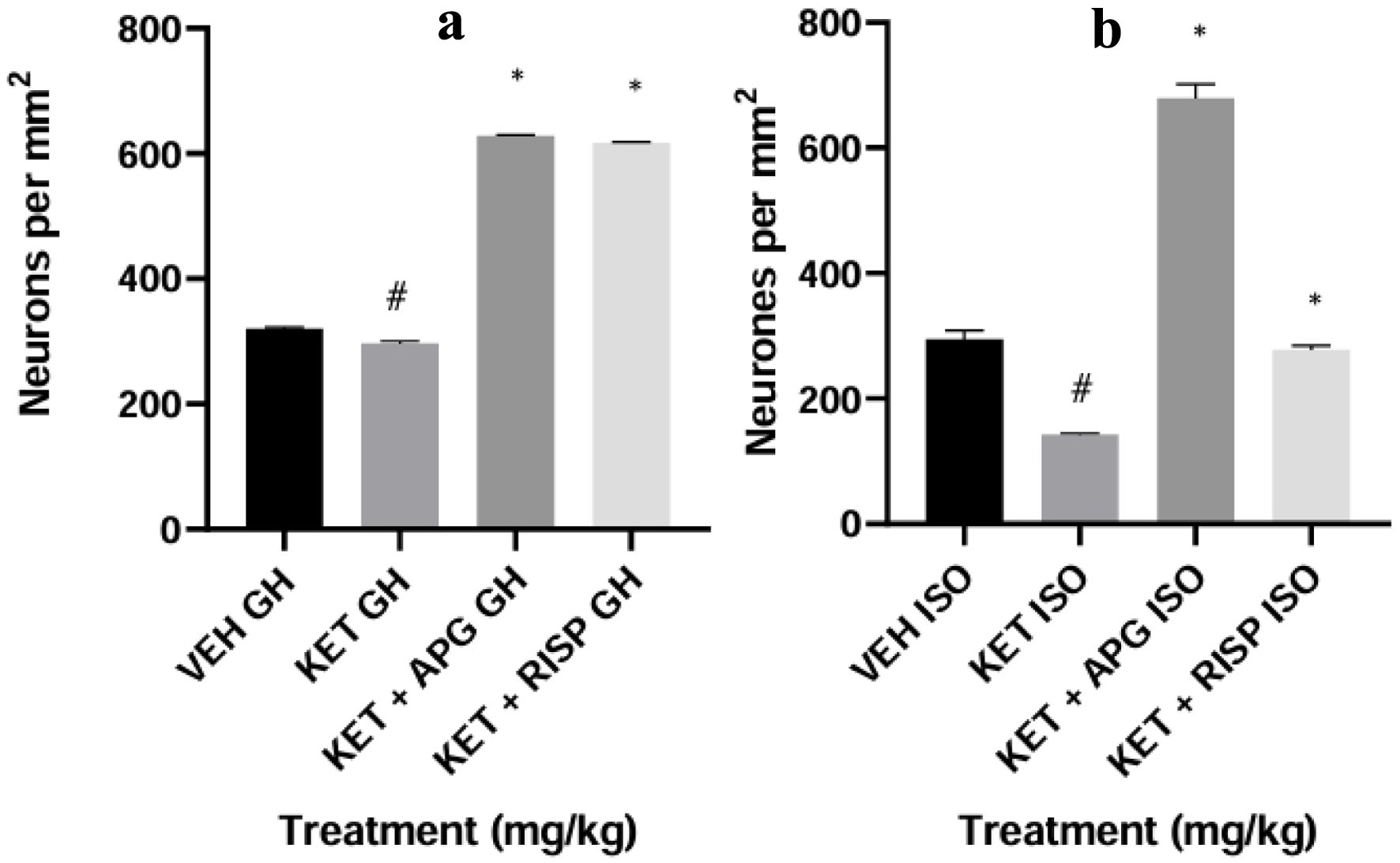 Click for large image | Figure 24. Neuronal density in hippocampus of weanling rats subjected to Two-Hit model of neurodevelopmental psychosis. (a) Group-housed animals. (b) Isolated-rearing animals. Bar represents the mean ± SEM of eight animals. *P < 0.05 as compared with KET GH/KET ISO followed by ANOVA and Tukey’s post hoc test for multiple comparisons. #P < 0.05 as compared with VEH GH/VEH ISO followed by ANOVA and Tukey’s post hoc test for multiple comparisons. APG: apigenin; ANOVA: analysis of variance; GH: group-housed; ISO: isolated; KET: ketamine; RISP: risperidone; SEM: standard error of the mean; VEH: vehicle. |
Histological assessment (H&E staining) of the PFC of weanling rats in two-hit model of neurodevelopmental psychosis
Photomicrographs portraying the histological and histomorphological (density of viable neuronal cells) using H&E staining demonstrated that administration of ketamine in group-housed post-weaned rats and isolated-rearing animals significantly (#P < 0.0001) showed cytoarchitectural changes in the PFC as evidenced in increased population of highly condensed (pyknotic) and angulated neuronal cells in relative to the group that received vehicle. More so, optical densitometry count showed that group that received ketamine in both group-housed animals and isolated-rearing animals showed significant reduction of viable neuronal cell population (*P < 0.0001) compared with apigenin and risperidone-treated group, as depicted in Figures 25 and 26.
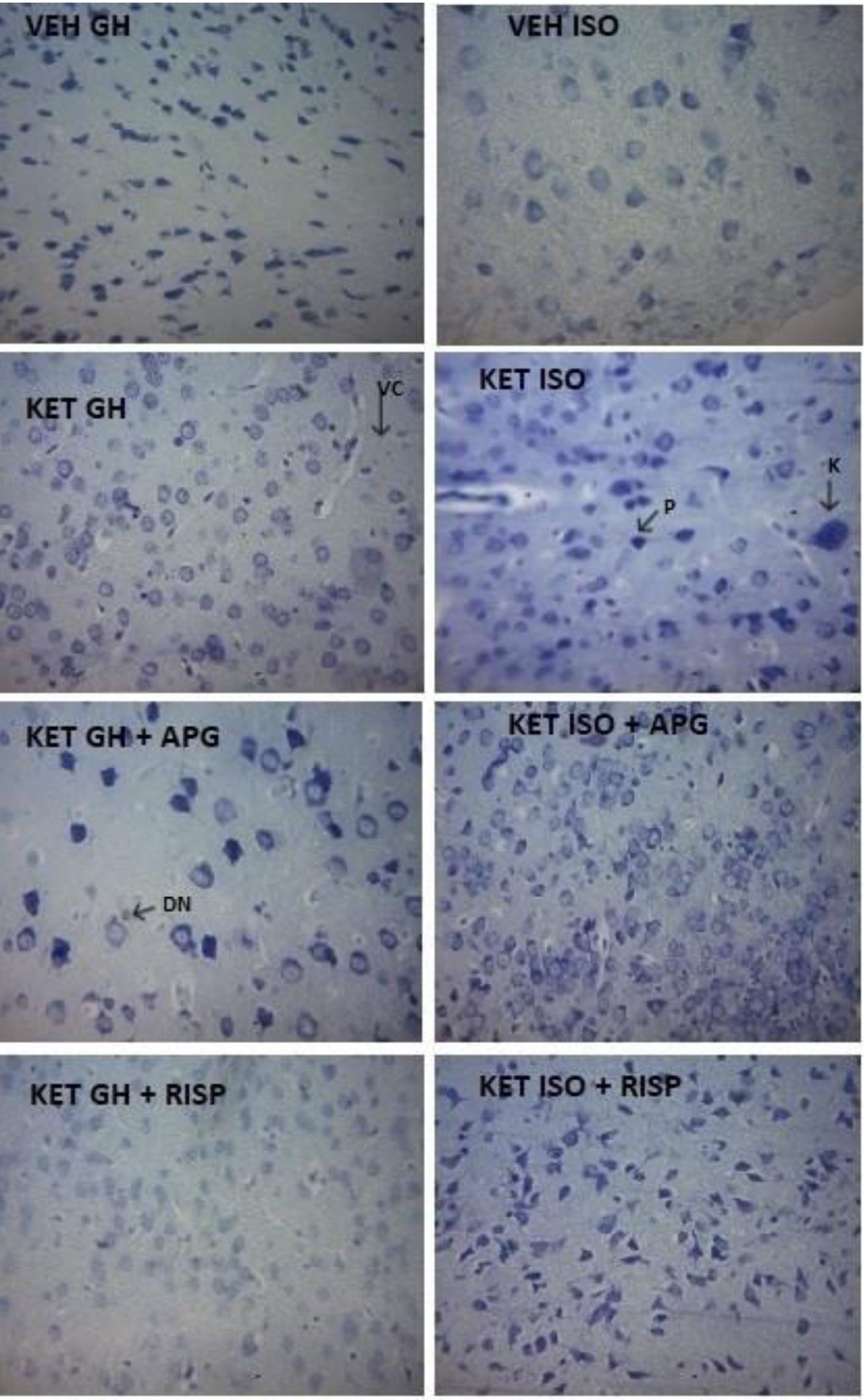 Click for large image | Figure 25. Photomicrographs of prefrontal cortex showing surviving neurons in weanling rats subjected to two-hit model of neurodevelopmental psychosis. APG: apigenin; DN: degenerating neurons; GH: group housed; ISO: isolated; K: karyopyknosis; KET: ketamine; P: pyknosis; RISP: risperidone; VC: vacoulated cytoplasm; VEH: vehicle. |
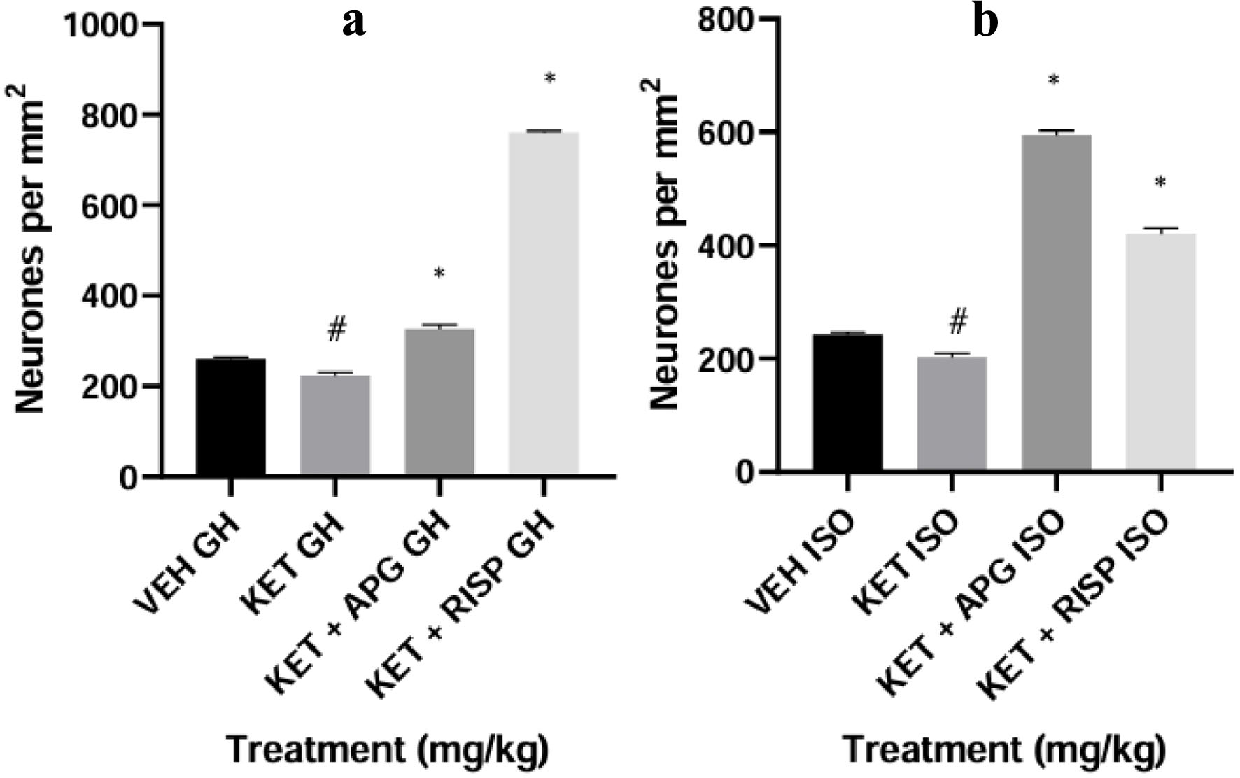 Click for large image | Figure 26. Neuronal density in prefrontal cortex of weanling rats subjected to two-hit model of neurodevelopmental psychosis. (a) Group-housed animals. (b) Isolated-rearing animals. Bar represents the mean ± SEM of eight animals. *P < 0.05 as compared with KET GH/KET ISO followed by ANOVA and Tukey’s post hoc test for multiple comparisons. #P < 0.05 as compared with VEH GH/VEH ISO followed by ANOVA and Tukey’s post hoc test for multiple comparisons. APG: apigenin; ANOVA: analysis of variance; GH: group-housed; ISO: isolated; KET: ketamine; RISP: risperidone; SEM: standard error of the mean; VEH: vehicle. |
| Discussion | ▴Top |
This study demonstrates the validity of two-hit neurodevelopmental model for evaluating the antipsychotic potential of novel compounds. The model combined early-life environmental stress post-weanling (social isolation rearing) with pharmacological induction of psychosis using ketamine, thereby mimicking both developmental and neurochemical components implicated in schizophrenia, aligning with evidence that early-life glutamatergic disruption alters neurodevelopmental processes implicated in schizophrenia [4, 6]. A total of 64 rats were assigned to eight experimental groups (n = 8 per group), consisting of four group-housed and four socially isolated cohorts. Animals were treated with ketamine, apigenin, or risperidone, allowing comparative assessment of behavioral, biochemical, neurochemical, and histological outcomes. Both group-housed and socially isolated rats exposed to ketamine exhibited pronounced schizophrenia-like behavioral alterations, confirming the validity of the model. These alterations were evident across behavioral paradigms and were consistent with previous reports demonstrating that early-life NMDA receptor antagonism disrupts neurodevelopmental trajectories. Such disruptions interfere with synaptogenesis, myelination, synaptic pruning, and axonal connectivity, processes known to be compromised in schizophrenia [47–50].
In the OFT, ketamine-treated group-housed rats showed significant hyperlocomotion, as reflected by increased number of line crossings compared with vehicle-treated controls. This hyperactivity is indicative of positive schizophrenia-like symptoms and dopaminergic dysregulation [25]. Notably, treatment with apigenin and risperidone markedly attenuated this hyperlocomotor activity. In contrast, chronically isolated rats displayed reduced locomotor activity following ketamine exposure, suggesting that prolonged social deprivation may exacerbate behavioral withdrawal and motivational deficits. Importantly, apigenin and risperidone normalized locomotor behavior in both housing conditions. The ability of apigenin to reverse locomotor abnormalities induced by the dual-hit model supports its putative antipsychotic-like properties, consistent with previous findings on apigenin with neuroactive potential [35].
Negative symptoms, particularly depression, represent a core and debilitating dimension of schizophrenia [51]. The FST was employed to model despair-like behavior, which has been widely accepted as a correlate of negative symptoms in rodents [52]. In this study, repeated administration of sub-anesthetic ketamine increased immobility time in group-housed rats, reflecting heightened behavioral despair and aligning with earlier reports linking ketamine exposure to depressive-like states [53]. Intervention with apigenin and risperidone significantly reduced immobility time in ketamine-treated group-housed rats. In socially isolated animals, ketamine alone did not significantly alter immobility compared to vehicle-treated isolated rats. However, apigenin and risperidone significantly reduced immobility duration, suggesting therapeutic efficacy even under compounded stress conditions. These findings indicate that apigenin, similar to risperidone, can alleviate negative symptoms of schizophrenia. The mechanistic basis for this effect may involve modulation of the serotonergic system, particularly the cortical 5-HT2A receptors [54]. Enhanced serotonergic tone has been implicated in the development of negative symptoms [55], and atypical antipsychotics such as risperidone and clozapine exert their effects partly through 5-HT2A antagonism, thereby mitigating ketamine-induced behavioral disturbances [56]. The comparable efficacy of apigenin suggests functional similarity to atypical antipsychotics in managing negative symptomatology.
Oxidative stress is a well-established contributor to schizophrenia pathophysiology [12]. In this study, key oxidative and nitrergic stress markers, GSH, SOD, MDA, and NO were evaluated in the amygdala, hippocampus, and PFC. Ketamine administration resulted in region-specific reductions in GSH and SOD, alongside elevations in MDA and NO levels, indicating enhanced lipid peroxidation and nitric oxide overproduction. These changes were particularly pronounced in group-housed ketamine-treated animals but were also evident under social isolation conditions. Intervention with apigenin and risperidone consistently restored antioxidant defenses, increased GSH and SOD levels, and reduced MDA and NO concentrations across all brain regions examined.
These findings confirm earlier reports that atypical antipsychotics such as risperidone enhance antioxidant capacity by increasing GSH and SOD while reducing NO levels [57]. The ability of apigenin to similarly reverse ketamine-induced oxidative imbalance underscores its neuroprotective and antioxidant properties, which likely contribute to its behavioral and neurochemical effects. apigenin, like vitamin C, is relatively an oxidant in comparison with GSH on the redox scale, and shows more positive values than the highest reported redox potential for GSH coupled to its oxidized partner, glutathione disulfide (GSSG) of −165 mV [58]. Thus, apigenin can be considered to be pro-oxidant when related to GSH [59]. This might explain difference in the scale of restoration of GSH antioxidant effects in the different brain sections. Future works, studying the direct interaction between apigenin and GSH utilizing assay that measures sulfhydryl levels, could provide more explanation. apigenin induces increased or preserved SOD expression at the messenger ribonucleic acid (mRNA) or protein level enhancing its antioxidant capacity in psychosis [60].
Furthermore, the present findings align with Estaphan et al 2021, who demonstrated that combined ketamine and social isolation significantly increased MDA levels and reduced antioxidant enzyme activity in the hippocampus and cortex [61]. Apigenin’s capacity to abate oxidative stress in the amygdala, hippocampus, and PFC further supports its therapeutic potential in neurodevelopmental psychosis.
Acetylcholine plays a critical role in cognition and memory, functions that are severely impaired in schizophrenia. Chronic ketamine exposure has been shown to disrupt cholinergic neurotransmission [23]. In the present study, AChE activity was increased in the PFC and amygdala of ketamine-treated group-housed rats, an effect reversed by apigenin and risperidone. This suggests enhanced cortical cholinergic signaling following treatment. In socially isolated rats, ketamine reduced AChE activity in the PFC and amygdala, and treatment with apigenin and risperidone further decreased AChE levels. In the hippocampus, AChE activity was reduced across both housing conditions following ketamine exposure and further attenuated by apigeninG and risperidone. Overall, apigenin’s ability to modulate AChE activity indicates its potential to attenuate ketamine-induced cognitive and memory dysfunction.
Dopaminergic dysregulation remains central to schizophrenia pathology [62]. Ketamine treatment reduced DA levels in the amygdala, hippocampus, and PFC of group-housed rats, effects that were reversed by apigenin and risperidone. Although no significant DA changes were observed in ketamine-treated isolated rats relative to vehicle controls, both apigenin and risperidone increased DA levels across all brain regions examined. It is a known fact that acute ketamine administration potentiates DA release in the brain, whereas chronic administration of ketamine decreases DA release in the brain region. apigenin increased DA levels in the brain regions, thus restoring the negative symptoms associated with psychosis [63]. These findings suggest that apigenin exerts dopaminergic modulatory effects, contributing to its antipsychotic-like profile.
Histological analysis revealed that chronic ketamine exposure significantly reduced viable neuronal cell counts in the amygdala, hippocampus, and PFC, accompanied by marked cytoarchitectural abnormalities, including increased pyknotic and degenerated neurons. Treatment with apigenin and risperidone preserved neuronal architecture and reduced degenerative changes, further supporting the neuroprotective role of flavonoids likely mediated through its antioxidant properties, which mitigate ketamine-induced ROS generation [64]. In conclusion, apigenin demonstrated antipsychotic-like, antioxidant, neurochemical, and neuroprotective effects comparable to risperidone in a two-hit model of schizophrenia. These findings highlight apigenin as a promising candidate for further investigation in the management of schizophrenia, particularly in addressing negative symptoms and oxidative stress-related pathology.
Apigenin like other flavonoid compounds could possess anti-inflammatory properties by mitigating the effects of pro-inflammatory cytokines, and also upregulate the expression of certain neurotrophic factors (brain-derived neurotrophic factor and nuclear factor-κB) as probable mechanisms of its antipsychotic effects.
Acknowledgments
The authors appreciate the supporting staff of the Department of Pharmacology and Therapeutics, Faculty of Basic Medical Sciences, University of Ibadan for the assistance rendered through the duration of the study.
Financial Disclosure
None to declare.
Conflict of Interest
The authors declare they have no competing interests.
Informed Consent
Not applicable.
Author Contributions
Conceptualization: Adegbuyi Oladele Aderibigbe, Adeniyi Nicholas Adesoye, Adewale Ganiyu Bakre. Investigation: Adegbuyi Oladele Aderibigbe, Adeniyi Nicholas Adesoye, Adewale Ganiyu Bakre, Wakeel Adegoke, Rotimi Olatunde. Methodology: Adegbuyi Oladele Aderibigbe, Adeniyi Nicholas Adesoye, Yusuf Usman, Adewale Ganiyu Bakre, Wakeel Adegoke, Rotimi Olatunde. Formal analysis: Adegbuyi Oladele Aderibigbe, Adeniyi Nicholas Adesoye, Adewale Ganiyu Bakre. Writing – original draft: Adeniyi Nicholas Adesoye, Yusuf Usman. Writing – review & editing: Adegbuyi Oladele Aderibigbe, Adewale Ganiyu Bakre. Supervision: Adegbuyi Oladele Aderibigbe, Adewale Ganiyu Bakre.
Data Availability
The authors declare that data supporting the findings of this study are available within the article.
Abbreviations
AChE: acetylcholinesterase; APG: apigenin; BDNF: brain-derived neurotrophic factor; DA: dopamine; DN: degenerating neurons; FST: forced swim test; GH: group-housed; GHK: group housed + ketamine; GHKAPG: group-housed, ketamine + apigenin; GHKRISP: group-housed, ketamine + risperidone; GSH: reduced glutathione; GSSG: glutathione disulphide; ISK: isolated + ketamine; ISO: isolated; ISV: isolated vehicle; K: karyopyknosis; KET: ketamine; MDA: malondialdehyde; NO: nitric oxide; OFT: open field test; P: pyknosis; PFC: prefrontal cortex; PND: postnatal day; RISP: risperidone; RNS: reactive nitrogen species; ROS: reactive oxygen species; SIKAPG: social isolation, ketamine + apigenin; SIKRISP: social isolation, ketamine + risperidone; SOD: superoxide dismutase; VC: vacoulated cytoplasm; VEH: vehicle
| References | ▴Top |
- Marder SR, Cannon TD. Schizophrenia. N Engl J Med. 2019;381(18):1753-1761.
doi pubmed - Jauhar S, Johnstone M, McKenna PJ. Schizophrenia. Lancet. 2022;399(10323):473-486.
doi pubmed - Patel KR, Cherian J, Gohil K, Atkinson D. Schizophrenia: overview and treatment options. P T. 2014;39(9):638-645.
pubmed - Martinez-Pinteno A, Rodriguez N, Olivares D, Madero S, Gomez M, Prohens L, Garcia-Rizo C, et al. Early treatment with JNJ-46356479, a mGluR2 modulator, improves behavioral and neuropathological deficits in a postnatal ketamine mouse model of schizophrenia. Biomed Pharmacother. 2023;158:114079.
doi pubmed - Guerrin CGJ, Doorduin J, Sommer IE, de Vries EFJ. The dual hit hypothesis of schizophrenia: Evidence from animal models. Neurosci Biobehav Rev. 2021;131:1150-1168.
doi pubmed - Shangase KB, Luvuno M, Mabandla M. Effects of combined postweaning social isolation and ketamine administration on schizophrenia-like behaviour in male Sprague Dawley rats. Behav Brain Res. 2025;476:115214.
doi pubmed - Graham B, James HM. Schizophrenia. Medicine.2024;52(8):476-480.
doi - Singh S, Khanna D, Kalra S. Role of neurochemicals in schizophrenia. Curr Psychopharmacol. 2020;9(2).144-161.
doi - Schmitt A, Falkai P, Papiol S. Neurodevelopmental disturbances in schizophrenia: evidence from genetic and environmental factors. J Neural Transm (Vienna). 2023;130(3):195-205.
doi pubmed - Petkovic A, Chaudhury D. Encore: Behavioural animal models of stress, depression and mood disorders. Front Behav Neurosci. 2022;16:931964.
doi pubmed - Herzberg MP, Gunnar MR. Early life stress and brain function: activity and connectivity associated with processing emotion and reward. Neuroimage. 2020;209:116493.
doi pubmed - Murray AJ, Rogers JC, Katshu M, Liddle PF, Upthegrove R. Oxidative stress and the pathophysiology and symptom profile of schizophrenia spectrum disorders. Front Psychiatry. 2021;12:703452.
doi pubmed - Zhang K, Zou L, Cai Y. Progress in the study of oxidative stress damage in patients with schizophrenia: challenges and opportunities. Front Psychiatry. 2025;16:1505397.
doi pubmed - Ermakov EA, Dmitrieva EM, Parshukova DA, Kazantseva DV, Vasilieva AR, Smirnova LP. Oxidative stress-related mechanisms in schizophrenia pathogenesis and new treatment perspectives. Oxid Med Cell Longev. 2021;2021:8881770.
doi pubmed - Hakenova K, Mikulecka A, Holubova K, Chvojkova M, Slamberova R, Jurcovicova J, Cechova B, et al. A new two-hit animal model for schizophrenia research: Consequences on social behavior. IBRO Neurosci Rep. 2025;19:38-49.
doi pubmed - Hany M, Rizvi A. Schizophrenia. In: StatPearls. Treasure Island (FL) ineligible companies. 2026.
pubmed - Pae CU. Role of the cholinesterase inhibitors in the treatment of schizophrenia. Expert Opin Investig Drugs. 2013;22(3):293-298.
doi pubmed - Saint-Georges Z, MacDonald J, Al-Khalili R, Hamati R, Solmi M, Keshavan MS, Tuominen L, et al. Cholinergic system in schizophrenia: a systematic review and meta-analysis. Mol Psychiatry. 2025;30(7):3301-3315.
doi pubmed - Tzavara ET, Bymaster FP, Davis RJ, Wade MR, Perry KW, Wess J, McKinzie DL, et al. M4 muscarinic receptors regulate the dynamics of cholinergic and dopaminergic neurotransmission: relevance to the pathophysiology and treatment of related CNS pathologies. FASEB J. 2004;18(12):1410-1412.
doi pubmed - Tandon R, Greden JF. Cholinergic hyperactivity and negative schizophrenic symptoms. A model of cholinergic/dopaminergic interactions in schizophrenia. Arch Gen Psychiatry. 1989;46(8):745-753.
doi pubmed - Acharya S, Kim KM. Roles of the functional interaction between brain cholinergic and dopaminergic systems in the pathogenesis and treatment of schizophrenia and Parkinson's disease. Int J Mol Sci. 2021;22(9):4299.
doi pubmed - Singh J, Kour K, Jayaram MB. Acetylcholinesterase inhibitors for schizophrenia. Cochrane Database Syst Rev. 2012;1(1):CD007967.
doi pubmed - Ben-Azu B, Adebayo OG, Fokoua AR, Oritsemuelebi B, Chidebe EO, Nwogueze CB, Kumanwee L, et al. Antipsychotic effect of diosgenin in ketamine-induced murine model of schizophrenia: Involvement of oxidative stress and cholinergic transmission. IBRO Neurosci Rep. 2024;16:86-97.
doi pubmed - Usman Y, Aderibigbe AO, Fehintola FA. Repeated ketamine doses elevate superoxide dismutase activity in a pharmacological model of schizophrenia-like phenotypes in mice. INNOSC Theranost & Pharm Sci. 2025.
doi - Usman Y, Aderibigbe AO, Ben-Azu B, Fehintola FA. Antipsychotic effects of ethanol extract of Blighia sapida (Sapindaecea) stem bark on pharmacological models of psychosis in Swiss mice. AJMMS. 2019;48:151-160.
- Uliana DL, Zhu X, Gomes FV, Grace AA. Using animal models for the studies of schizophrenia and depression: The value of translational models for treatment and prevention. Front Behav Neurosci. 2022;16:935320.
doi pubmed - Monte AS, Mello BSF, Borella VCM, da Silva Araujo T, da Silva FER, Sousa FCF, de Oliveira ACP, et al. Two-hit model of schizophrenia induced by neonatal immune activation and peripubertal stress in rats: Study of sex differences and brain oxidative alterations. Behav Brain Res. 2017;331:30-37.
doi pubmed - Guma E, Cupo L, Ma W, Gallino D, Moquin L, Gratton A, Devenyi GA, et al. Investigating the "two-hit hypothesis": Effects of prenatal maternal immune activation and adolescent cannabis use on neurodevelopment in mice. Prog Neuropsychopharmacol Biol Psychiatry. 2023;120:110642.
doi pubmed - Ram E, Raphaeli S, Avital A. Prepubertal chronic stress and ketamine administration to rats as a neurodevelopmental model of schizophrenia symptomatology. Int J Neuropsychopharmacol. 2013;16(10):2307-2314.
doi pubmed - Kramer DJ, Johnson AA. Apigenin: a natural molecule at the intersection of sleep and aging. Front Nutr. 2024;11:1359176.
doi pubmed - Allemailem KS, Almatroudi A, Alharbi HOA, AlSuhaymi N, Alsugoor MH, Aldakheel FM, Khan AA, et al. Apigenin: a bioflavonoid with a promising role in disease prevention and treatment. Biomedicines. 2024;12(6):1353.
doi pubmed - Patil RH, Babu RL, Naveen Kumar M, Kiran Kumar KM, Hegde SM, Nagesh R, Ramesh GT, et al. Anti-inflammatory effect of apigenin on LPS-induced pro-inflammatory mediators and AP-1 factors in human lung epithelial cells. Inflammation. 2016;39(1):138-147.
doi pubmed - Liu R, Zhang T, Yang H, Lan X, Ying J, Du G. The flavonoid apigenin protects brain neurovascular coupling against amyloid-beta(2)(5)(-)(3)(5)-induced toxicity in mice. J Alzheimers Dis. 2011;24(1):85-100.
doi pubmed - Nabavi SF, Khan H, D'Onofrio G, Samec D, Shirooie S, Dehpour AR, Arguelles S, et al. Apigenin as neuroprotective agent: Of mice and men. Pharmacol Res. 2018;128:359-365.
doi pubmed - Oyagbemi AA, Femi-Akinlosotu OM, Obasa AA, Ojo MS, Salami AT, Ajibade TO, Onukak CE, et al. Apigenin mitigates oxidative stress, neuroinflammation, and cognitive impairment but enhances learning and memory in aluminum chloride-induced neurotoxicity in rats. Alzheimers Dement. 2025;21(5):e70223.
doi pubmed - Koh MT, Shao Y, Sherwood A, Smith DR. Impaired hippocampal-dependent memory and reduced parvalbumin-positive interneurons in a ketamine mouse model of schizophrenia. Schizophr Res. 2016;171(1-3):187-194.
doi pubmed - Monte AS, de Souza GC, McIntyre RS, Soczynska JK, dos Santos JV, Cordeiro RC, Ribeiro BM, et al. Prevention and reversal of ketamine-induced schizophrenia related behavior by minocycline in mice: Possible involvement of antioxidant and nitrergic pathways. J Psychopharmacol. 2013;27(11):1032-1043.
doi pubmed - Chindo BA, Adzu B, Yahaya TA, Gamaniel KS. Ketamine-enhanced immobility in forced swim test: a possible animal model for the negative symptoms of schizophrenia. Prog Neuropsychopharmacol Biol Psychiatry. 2012;38(2):310-316.
doi pubmed - Kroczka B, Branski P, Palucha A, Pilc A, Nowak G. Antidepressant-like properties of zinc in rodent forced swim test. Brain Res Bull. 2001;55(2):297-300.
doi pubmed - Weiner I, Schiller D, Gaisler-Salomon I, Green A, Joel D. A comparison of drug effects in latent inhibition and the forced swim test differentiates between the typical antipsychotic haloperidol, the atypical antipsychotics clozapine and olanzapine, and the antidepressants imipramine and paroxetine. Behav Pharmacol. 2003;14(3):215-222.
doi pubmed - Moron MS, Depierre JW, Mannervik B. Levels of glutathione, glutathione reductase and glutathione S-transferase activities in rat lung and liver. Biochim Biophys Acta. 1979;582(1):67-78.
doi pubmed - Ohkawa H, Ohishi N, Yagi K. Assay for lipid peroxides in animal tissues by thiobarbituric acid reaction. Anal Biochem. 1979;95(2):351-358.
doi pubmed - Green LC, Wagner DA, Glogowski J, Skipper PL, Wishnok JS, Tannenbaum SR. Analysis of nitrate, nitrite, and [15N]nitrate in biological fluids. Anal Biochem. 1982;126(1):131-138.
doi pubmed - Misra HP, Fridovich I. The role of superoxide anion in the autoxidation of epinephrine and a simple assay for superoxide dismutase. J Biol Chem. 1972;247(10):3170-3175.
pubmed - Ellman GL, Courtney KD, Andres V, Jr., Feather-Stone RM. A new and rapid colorimetric determination of acetylcholinesterase activity. Biochem Pharmacol. 1961;7:88-95.
doi pubmed - Gornall AG, Bardawill CJ, David MM. Determination of serum proteins by means of the biuret reaction. J Biol Chem. 1949;177(2):751-766.
pubmed - Babikian T, Satz P, Zaucha K, Light R, Lewis RS, Asarnow RF. The UCLA longitudinal study of neurocognitive outcomes following mild pediatric traumatic brain injury. J Int Neuropsychol Soc. 2011;17(5):886-895.
doi pubmed - Chini M, Pfeffer T, Hanganu-Opatz I. An increase of inhibition drives the developmental decorrelation of neural activity. Elife. 2022;11.
doi pubmed - Germann M, Brederoo SG, Sommer IEC. Abnormal synaptic pruning during adolescence underlying the development of psychotic disorders. Curr Opin Psychiatry. 2021;34(3):222-227.
doi pubmed - Osimo EF, Baxter LJ, Lewis G, Jones PB, Khandaker GM. Prevalence of low-grade inflammation in depression: a systematic review and meta-analysis of CRP levels. Psychol Med. 2019;49(12):1958-1970.
doi pubmed - Correll CU, Schooler NR. Negative symptoms in schizophrenia: a review and clinical guide for recognition, assessment, and treatment. Neuropsychiatr Dis Treat. 2020;16:519-534.
doi pubmed - Yankelevitch-Yahav R, Franko M, Huly A, Doron R. The forced swim test as a model of depressive-like behavior. J Vis Exp. 2015;97:e52587.
doi pubmed - Chang H, Huang MC, Chen LY. Major depressive disorder induced by chronic ketamine abuse: a case report. Prim Care Companion CNS Disord. 2016;18(3).
doi pubmed - Romeo B, Willaime L, Rari E, Benyamina A, Martelli C. Efficacy of 5-HT2A antagonists on negative symptoms in patients with schizophrenia: A meta-analysis. Psychiatry Res. 2023;321:115104.
doi pubmed - Osugo M, Whitehurst T, Erritzoe D, Carr R, Ashok AH, Maccioni L, Onwordi EC, et al. Role of serotonin in the neurobiology of schizophrenia and association with negative symptoms. JAMA Psychiatry. 2026;83(2):185-195.
doi pubmed - Kusumi I, Boku S, Takahashi Y. Psychopharmacology of atypical antipsychotic drugs: From the receptor binding profile to neuroprotection and neurogenesis. Psychiatry Clin Neurosci. 2015;69(5):243-258.
doi pubmed - Tendilla-Beltran H, Meneses-Prado S, Vazquez-Roque RA, Tapia-Rodriguez M, Vazquez-Hernandez AJ, Coatl-Cuaya H, Martin-Hernandez D, et al. Risperidone ameliorates prefrontal cortex neural atrophy and oxidative/nitrosative stress in brain and peripheral blood of rats with neonatal ventral hippocampus lesion. J Neurosci. 2019;39(43):8584-8599.
doi pubmed - Kirlin WG, Cai J, Thompson SA, Diaz D, Kavanagh TJ, Jones DP. Glutathione redox potential in response to differentiation and enzyme inducers. Free Radic Biol Med. 1999;27(11-12):1208-1218.
doi pubmed - Andueza A, Garcia-Garzon A, Ruiz de Galarreta M, Ansorena E, Iraburu MJ, Lopez-Zabalza MJ, Martinez-Irujo JJ. Oxidation pathways underlying the pro-oxidant effects of apigenin. Free Radic Biol Med. 2015;87:169-180.
doi pubmed - Zhou N, Tian Y, Liu W, Tu B, Xu W, Gu T, Zou K, et al. Protective Effects of Resveratrol and Apigenin Dietary Supplementation on Serum Antioxidative Parameters and mRNAs Expression in the Small Intestines of Diquat-Challenged Pullets. Front Vet Sci. 2022;9:850769.
doi pubmed - Estaphan S, Curpan AS, Khalifa D, Rashed L, Ciobica A, Cantemir A, Ciobica A, et al. Combined low dose of ketamine and social isolation: a possible model of induced chronic schizophrenia-like symptoms in male albino rats. Brain Sci. 2021;11(7).
doi pubmed - Kokkinou M, Irvine EE, Bonsall DR, Natesan S, Wells LA, Smith M, Glegola J, et al. Reproducing the dopamine pathophysiology of schizophrenia and approaches to ameliorate it: a translational imaging study with ketamine. Mol Psychiatry. 2021;26(6):2562-2576.
doi pubmed - Kokkinou M, Ashok AH, Howes OD. The effects of ketamine on dopaminergic function: meta-analysis and review of the implications for neuropsychiatric disorders. Mol Psychiatry. 2018;23(1):59-69.
doi pubmed - Ishola IO, Ben-Azu B, Adebayo OA, Ajayi AM, Omorodion IL, Edje KE, Adeyemi OO. Prevention and reversal of ketamine-induced experimental psychosis in mice by the neuroactive flavonoid, hesperidin: The role of oxidative and cholinergic mechanisms. Brain Res Bull. 2021;177:239-251.
doi pubmed
This article is distributed under the terms of the Creative Commons Attribution 4.0 International License (CC BY 4.0), which permits unrestricted use, distribution, and reproduction in any medium, including commercial use, provided the original work is properly cited.
Journal of Current Pharmacology is published by Elmer Press Inc.